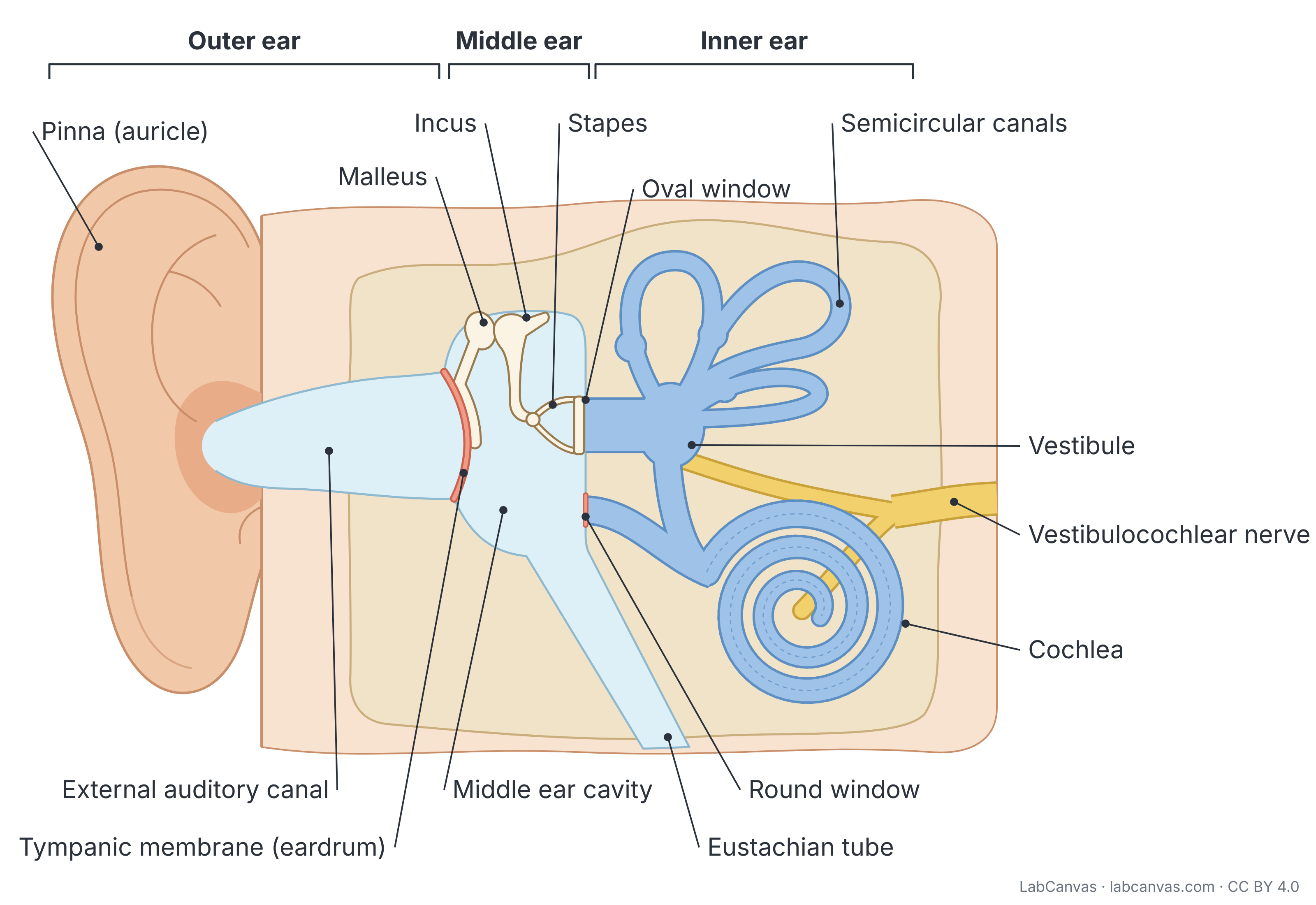
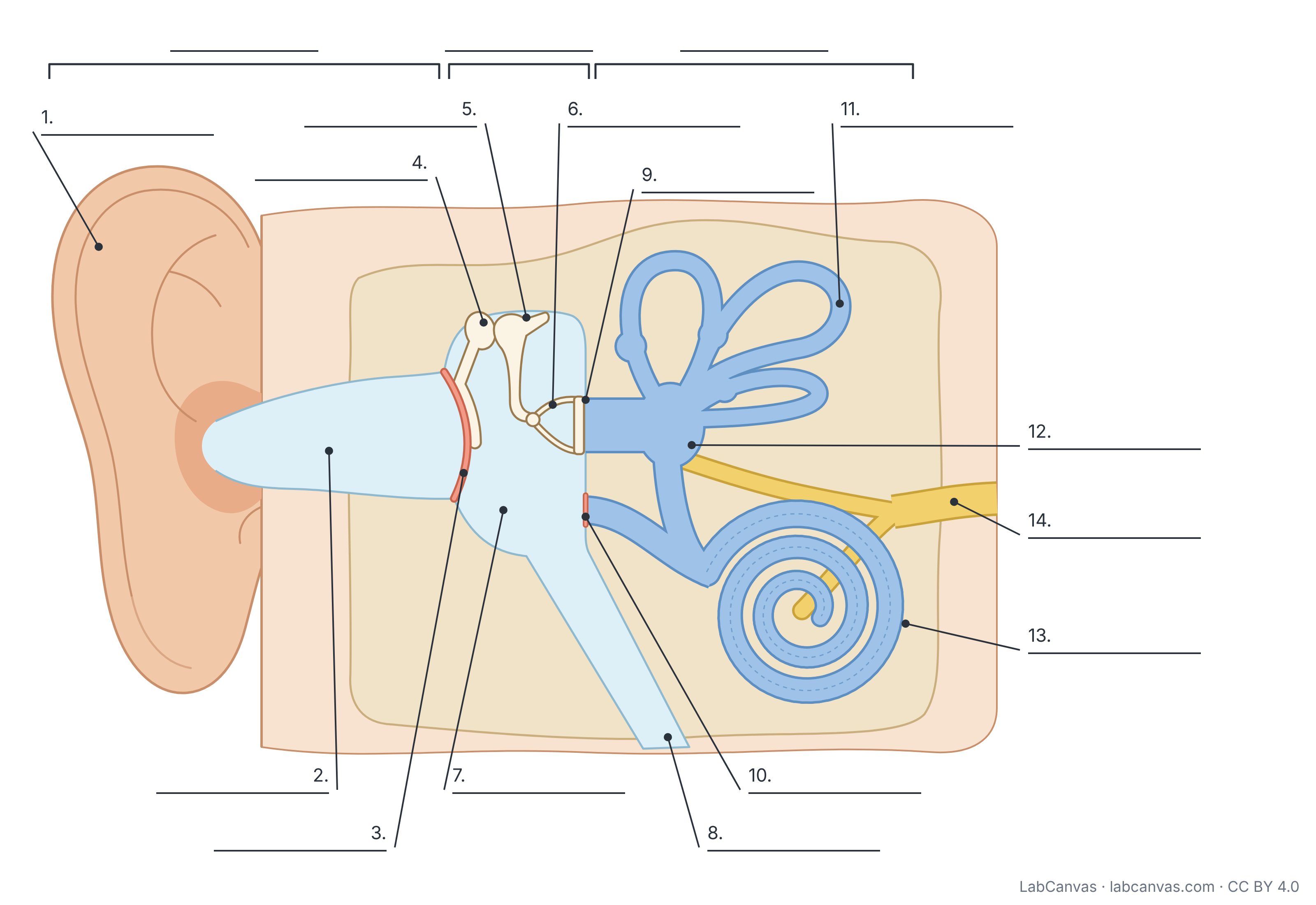
Ear diagram (labelled parts of the human ear)
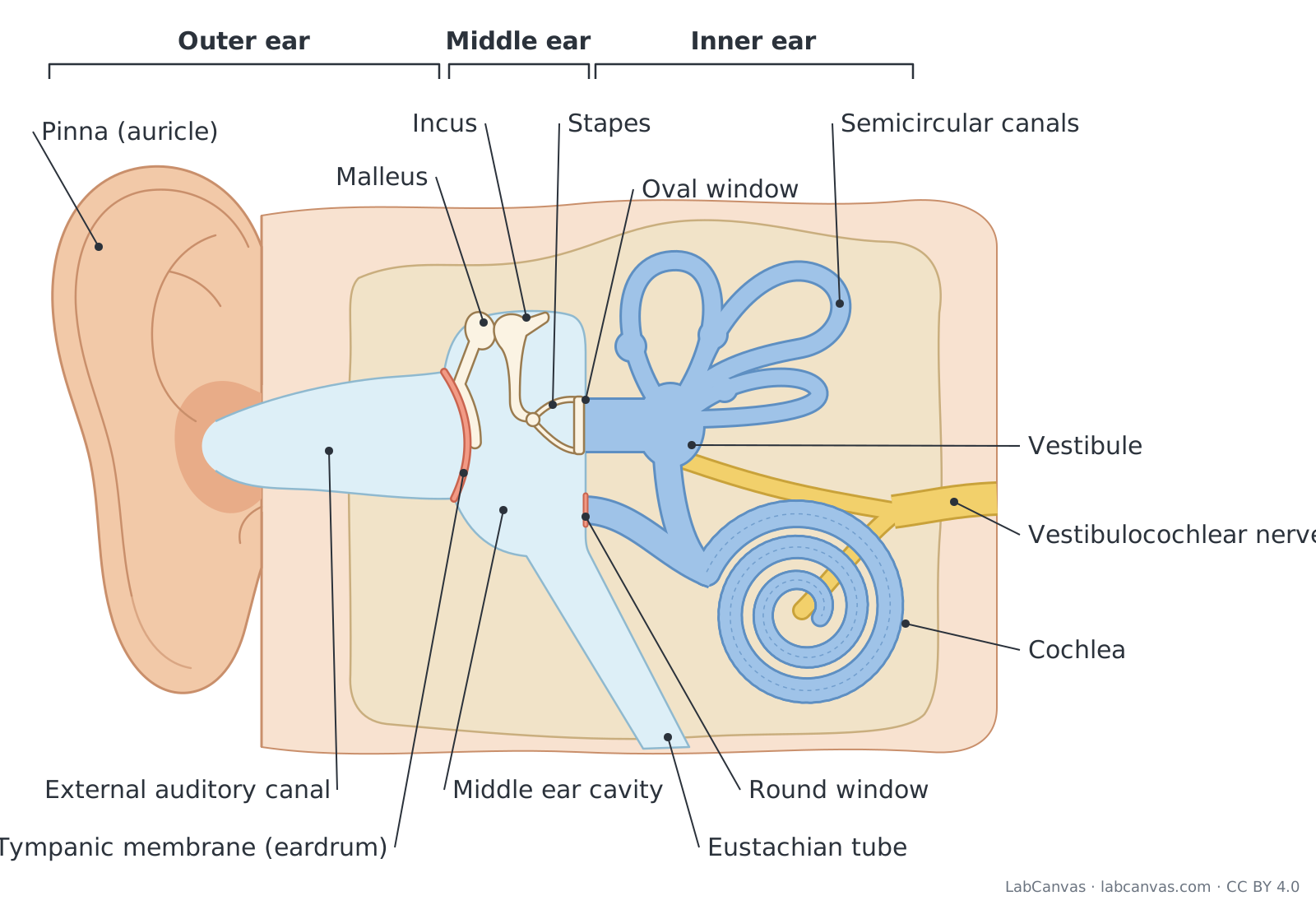
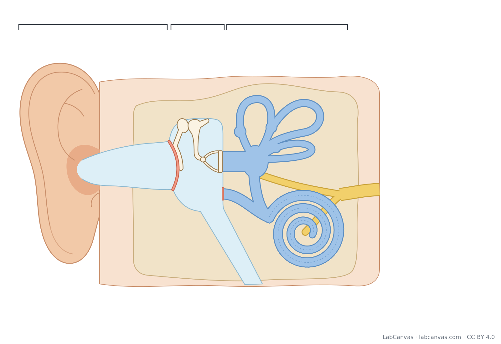
The human ear has three parts. The outer ear (pinna and external auditory canal) collects sound and channels it to the tympanic membrane (eardrum). The middle ear is an air-filled cavity where three tiny bones, the malleus, incus and stapes, carry the eardrum’s vibrations to the oval window, and the Eustachian tube links it to the throat. The fluid-filled inner ear holds the cochlea, which turns vibrations into nerve signals for hearing, and the vestibule and semicircular canals, which sense balance; the vestibulocochlear nerve carries both to the brain.
Last updated
Download the ear diagram
Free to use in homework, teaching, slides, posters and publications under CC BY 4.0. Please credit: Ear diagram by LabCanvas (labcanvas.com), CC BY 4.0
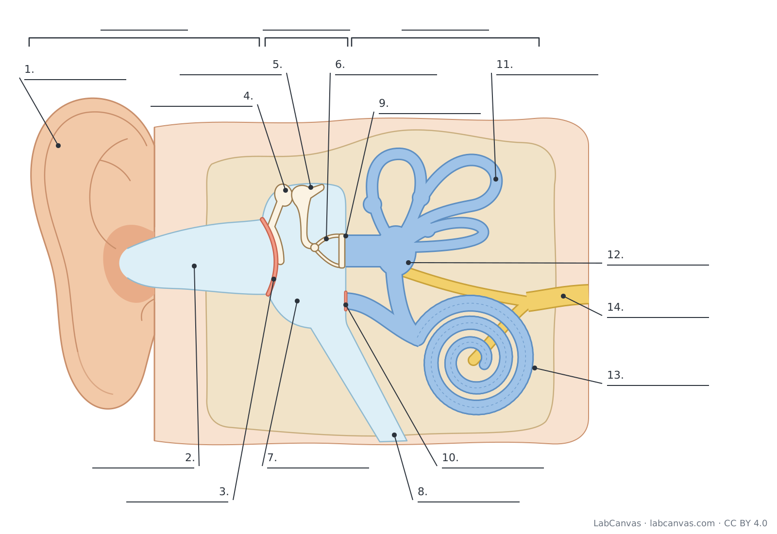
Parts of the ear and what they do
| # | Part | What it is or does |
|---|---|---|
| 1 | Pinna (auricle) | The visible flap of skin-covered cartilage that collects sound waves and funnels them into the ear canal; its shape also helps you tell where a sound comes from. |
| 2 | External auditory canal | The ear canal: a tube that carries sound from the pinna to the eardrum; glands in its lining make earwax that traps dust. |
| 3 | Tympanic membrane (eardrum) | A thin membrane across the end of the ear canal that vibrates when sound waves hit it, separating the outer ear from the middle ear. |
| 4 | Malleus (hammer) | The first ossicle: its handle is attached to the eardrum and passes the vibrations on to the incus. |
| 5 | Incus (anvil) | The middle ossicle, linking the malleus to the stapes. |
| 6 | Stapes (stirrup) | The last and smallest ossicle (the smallest bone in the body); its footplate pushes on the oval window. |
| 7 | Middle ear cavity | The air-filled space in the temporal bone that holds the ossicles, between the eardrum and the inner ear. |
| 8 | Eustachian (auditory) tube | Connects the middle ear to the upper throat (nasopharynx) so the air pressure on both sides of the eardrum stays equal. |
| 9 | Oval window | A membrane-covered opening into the inner ear where the stapes footplate sits; its movement sets the inner-ear fluid in motion. |
| 10 | Round window | A second membrane-covered opening below the oval window that bulges outward as the fluid moves, letting the pressure wave escape. |
| 11 | Semicircular canals | Three fluid-filled loops set roughly at right angles to each other; they detect rotation of the head for balance. |
| 12 | Vestibule | The central chamber of the inner ear; its utricle and saccule sense head position relative to gravity and straight-line movement. |
| 13 | Cochlea | The snail-shaped, fluid-filled organ of hearing: hair cells on its basilar membrane turn vibrations into nerve signals. |
| 14 | Vestibulocochlear nerve | Cranial nerve VIII: its cochlear branch carries hearing signals and its vestibular branch carries balance signals to the brain. |
How to label a ear diagram
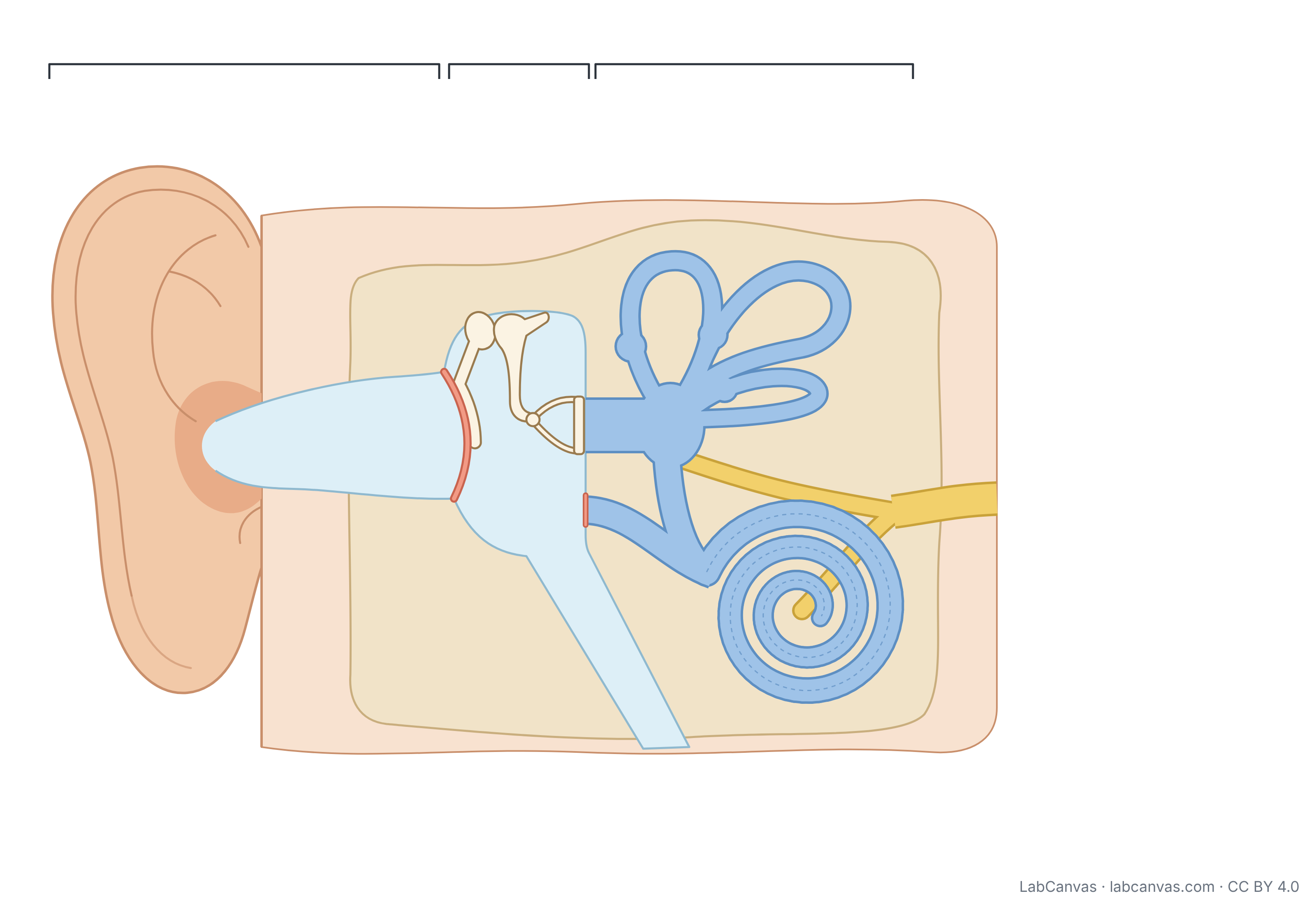
- Split the ear into three regionsBracket the outer ear (pinna to eardrum), the middle ear (eardrum to the oval window) and the inner ear (the cochlea, vestibule and semicircular canals). Lateral, the outside of the head, is usually drawn on the left.
- Label the outer earLabel the pinna (auricle), then follow the tube inwards and label the external auditory canal.
- Label the eardrumThe thin, slightly cone-shaped membrane closing the end of the canal is the tympanic membrane (eardrum).
- Label the ossicles in orderInside the middle ear cavity, label the malleus (touching the eardrum), the incus (in the middle) and the stapes (the stirrup in the oval window). A memory aid: hammer, anvil, stirrup.
- Label the openings and the tubeOn the inner wall of the middle ear, label the oval window (with the stapes) and the round window just below it. Label the Eustachian tube running down from the middle ear towards the throat.
- Label the inner earLabel the three loops of the semicircular canals, the vestibule they join, and the coiled cochlea.
- Finish with the nerveLabel the vestibulocochlear nerve leaving the cochlea and vestibule towards the brain, then check every leader line points to the right structure.
Outer, middle and inner ear compared
| Region | Parts | Filled with | Job |
|---|---|---|---|
| Outer ear | Pinna, external auditory canal, outer surface of the eardrum | Air | Collects sound waves and channels them to the eardrum |
| Middle ear | Tympanic membrane, malleus, incus, stapes, middle ear cavity, Eustachian tube | Air | Turns sound into vibrations of the ossicles and passes them, amplified, to the oval window; keeps air pressure equal |
| Inner ear | Oval and round windows, cochlea, vestibule, semicircular canals, vestibulocochlear nerve | Fluid | Cochlea converts vibrations into nerve signals (hearing); vestibule and semicircular canals sense balance |
How hearing works, step by step
- The pinna collects sound waves (pressure waves in the air) and funnels them down the external auditory canal.
- The sound waves make the tympanic membrane (eardrum) vibrate.
- The malleus, incus and stapes pass the vibration across the middle ear. Because the eardrum is much larger than the oval window, the force is concentrated, which amplifies the pressure enough to move fluid.
- The stapes rocks in the oval window and sends pressure waves through the fluid of the cochlea; the round window bulges outward to let the fluid move.
- The waves make the basilar membrane vibrate. Hair cells on it bend their stereocilia, which opens ion channels and triggers nerve impulses.
- The cochlear branch of the vestibulocochlear nerve carries the impulses to the brainstem and on to the auditory cortex, where they are perceived as sound.
Pitch depends on where the basilar membrane vibrates most: high-frequency sounds stimulate the stiff, narrow base of the cochlea and low-frequency sounds the wider apex. Louder sounds make the membrane vibrate more strongly, stimulating more hair cells.
How the ear controls balance
The vestibule and semicircular canals form the vestibular system, which shares the inner ear and nerve with hearing but is not involved in hearing. The three semicircular canals sit roughly at right angles to one another. When the head turns, the fluid inside lags behind and bends hair cells in the swelling at the base of each canal (the ampulla), signalling rotation. In the vestibule, the utricle and saccule contain hair cells under tiny crystals (otoliths) that shift with gravity and straight-line acceleration, signalling head position. These signals travel in the vestibular branch of the vestibulocochlear nerve.
Why ears “pop”
The middle ear is a closed air space. When the outside air pressure changes quickly, for example in a plane or a lift, the eardrum is pushed in or out and sounds become muffled. Swallowing or yawning opens the Eustachian tube, letting air in or out of the middle ear until the pressure on both sides of the eardrum is equal again: that is the “pop”.
Quick quiz
1. Which structure separates the outer ear from the middle ear?
- Oval window
- Tympanic membrane
- Round window
- Cochlea
Answer: b) Tympanic membrane. The tympanic membrane (eardrum) closes the inner end of the ear canal.
2. What is the correct order of the ossicles from the eardrum inwards?
- Stapes, incus, malleus
- Incus, malleus, stapes
- Malleus, incus, stapes
- Malleus, stapes, incus
Answer: c) Malleus, incus, stapes. The malleus touches the eardrum, the incus is in the middle and the stapes sits in the oval window.
3. Which part of the ear turns vibrations into nerve signals for hearing?
- Semicircular canals
- Eustachian tube
- Cochlea
- Pinna
Answer: c) Cochlea. Hair cells in the cochlea convert fluid vibrations into nerve impulses.
4. What does the Eustachian tube do?
- Carries sound to the brain
- Equalises air pressure in the middle ear
- Detects rotation of the head
- Produces earwax
Answer: b) Equalises air pressure in the middle ear. It links the middle ear to the throat so pressure on both sides of the eardrum stays equal.
5. The semicircular canals are mainly involved in…
- Hearing high-pitched sounds
- Balance, by sensing head rotation
- Amplifying sound
- Equalising pressure
Answer: b) Balance, by sensing head rotation. Fluid in the canals moves when the head turns, bending hair cells that signal rotation.
Frequently asked questions
What are the labels of the ear?
The main labels of a human ear diagram are: pinna (auricle), external auditory canal, tympanic membrane (eardrum), malleus, incus, stapes, middle ear cavity, Eustachian (auditory) tube, oval window, round window, semicircular canals, vestibule, cochlea and vestibulocochlear (auditory) nerve, grouped into the outer, middle and inner ear.
What are the three main parts of the ear?
The outer ear (pinna and ear canal), the middle ear (eardrum, the three ossicles and the Eustachian tube) and the inner ear (cochlea, vestibule and semicircular canals).
What are the three small bones in the ear called?
The ossicles: the malleus (hammer), incus (anvil) and stapes (stirrup). They carry vibrations from the eardrum to the oval window, and the stapes is the smallest bone in the human body.
What does the cochlea do?
The cochlea is the organ of hearing. Pressure waves in its fluid make the basilar membrane vibrate, bending the stereocilia of hair cells, which then trigger nerve impulses in the cochlear nerve.
Which part of the ear controls balance?
The inner ear’s vestibular system: the semicircular canals sense rotation of the head, and the utricle and saccule in the vestibule sense head position and straight-line movement.
What is the Eustachian tube for?
It connects the middle ear to the upper throat and opens when you swallow or yawn, keeping the air pressure in the middle ear equal to the pressure outside so the eardrum can vibrate freely.
How do you label a diagram of the ear?
Work from the outside in: pinna, ear canal and eardrum (outer ear); malleus, incus, stapes and the Eustachian tube (middle ear); oval and round windows, semicircular canals, vestibule, cochlea and the vestibulocochlear nerve (inner ear).
Sources
- OpenStax Biology 2e, 36.4 Hearing and Vestibular Sensation
- OpenStax Anatomy and Physiology 2e, 14.1 Sensory Perception (Hearing)
Facts on this page follow these introductory textbooks (OpenStax, CC BY 4.0). The diagram is a simplified illustration; organelles are not drawn to scale.
More free diagrams
Make your own science diagrams
LabCanvas is a browser workspace for scientific figures, slides and posters, with adjustable cells, organelles and molecules.
Edit this diagram