Cell membrane diagram (fluid mosaic model)
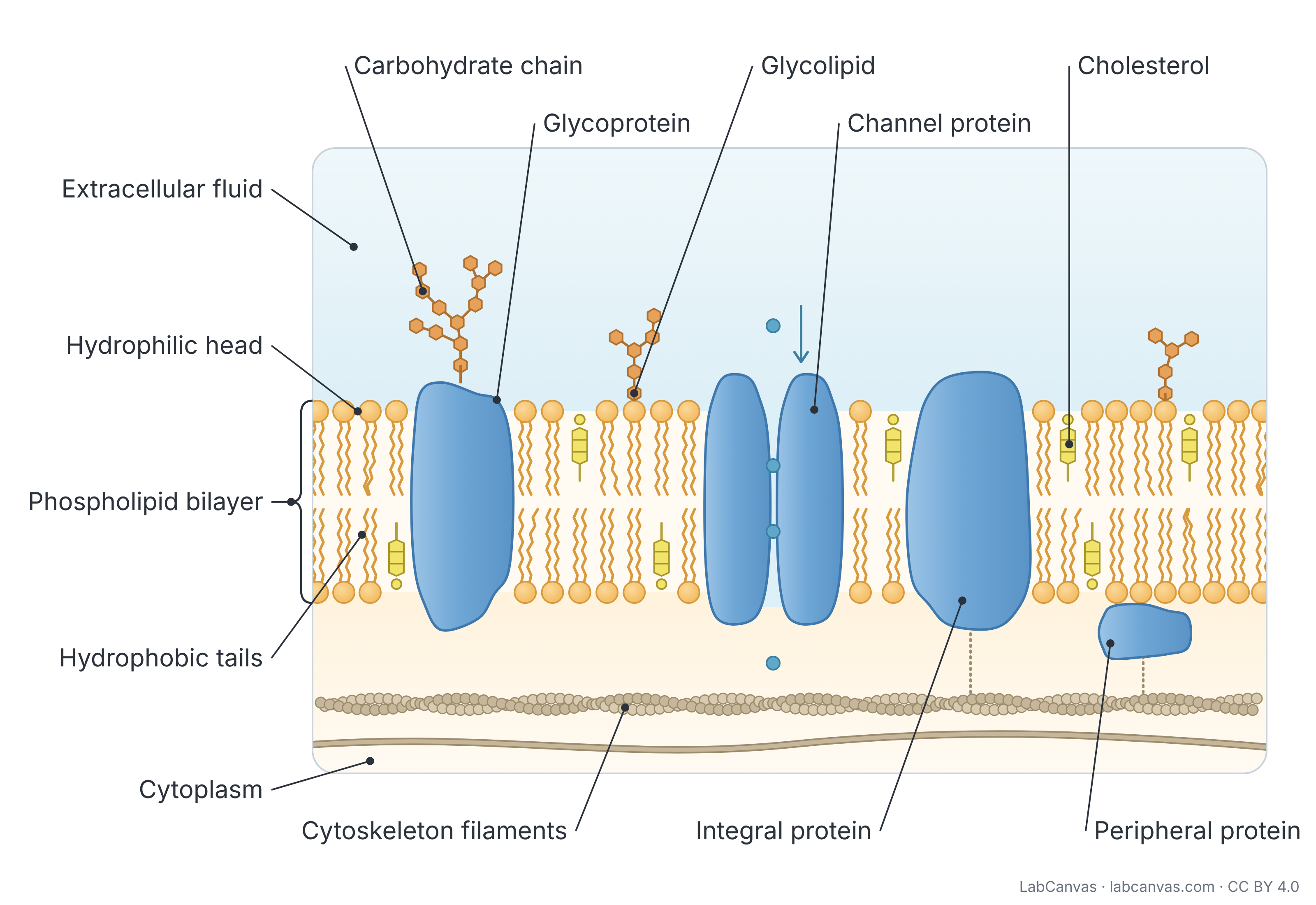
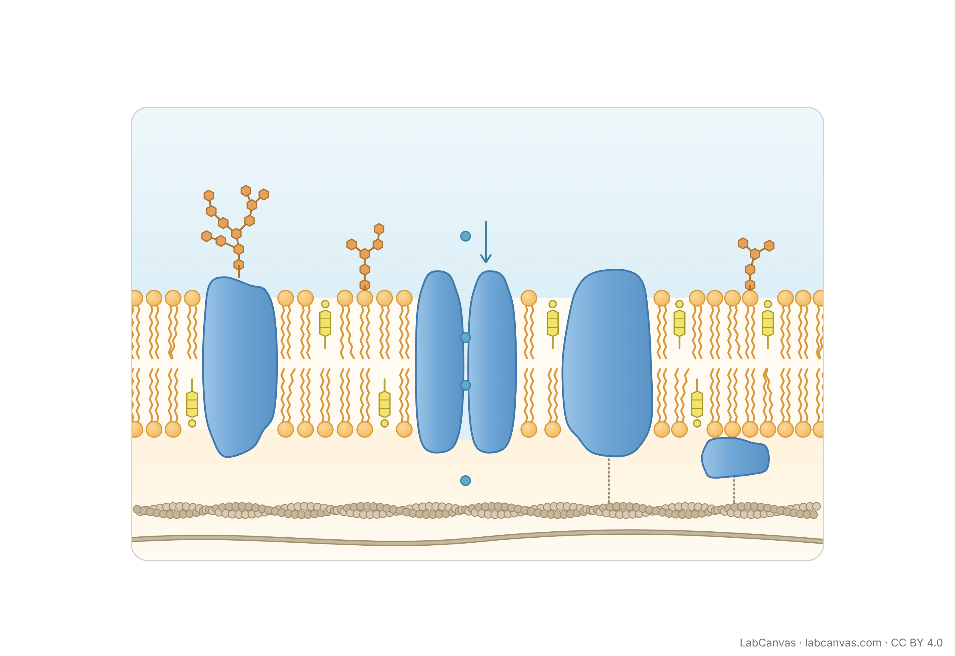
A cell membrane diagram shows the plasma membrane as a phospholipid bilayer: two layers of phospholipids with their hydrophilic heads facing the watery fluid on each side and their hydrophobic tails facing inward. Proteins sit in and on the bilayer, cholesterol is tucked between the phospholipids, and carbohydrate chains attached to proteins (glycoproteins) and lipids (glycolipids) face the extracellular fluid. Because the parts can move sideways and the proteins are scattered like tiles, this picture is called the fluid mosaic model.
Last updated
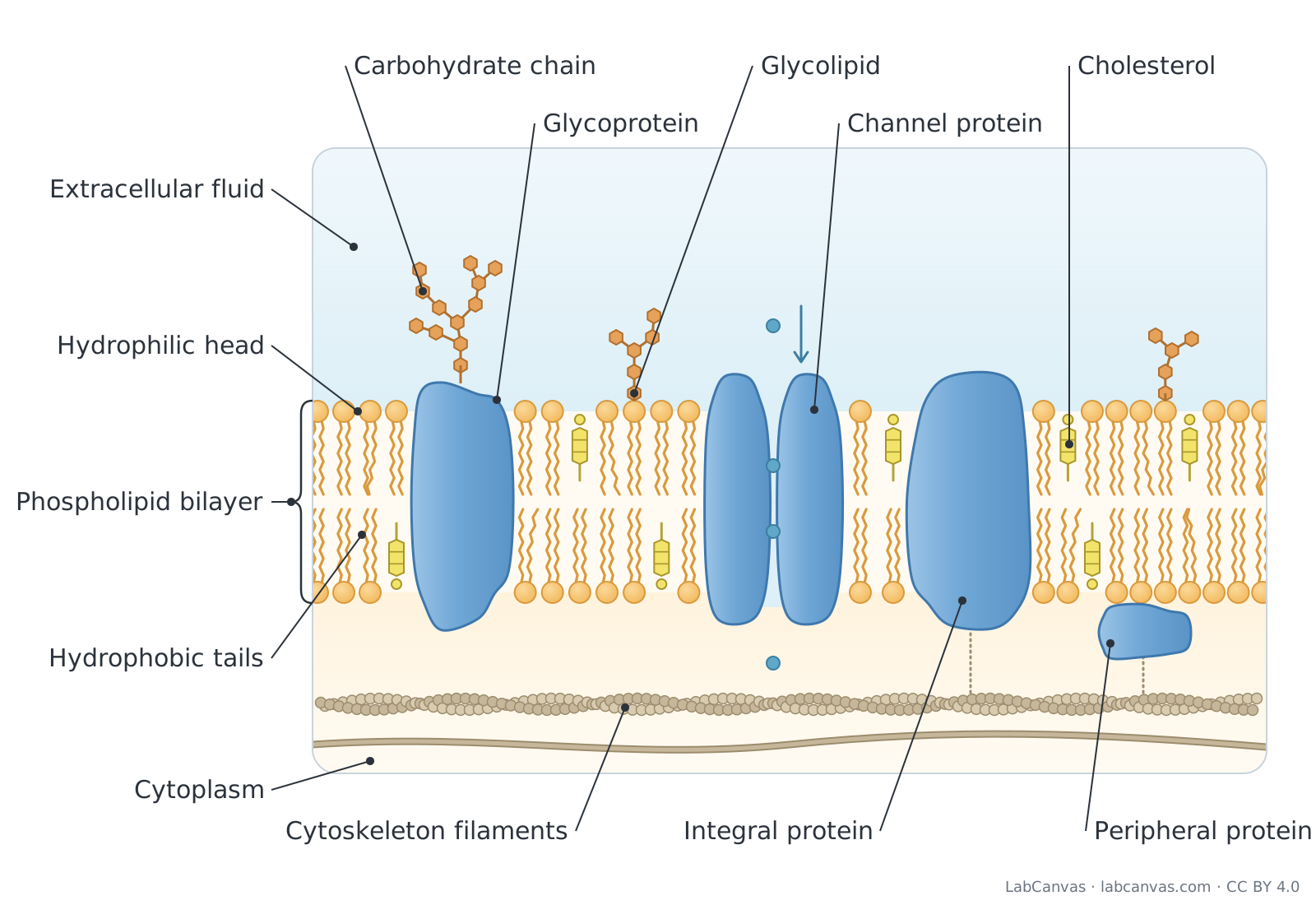
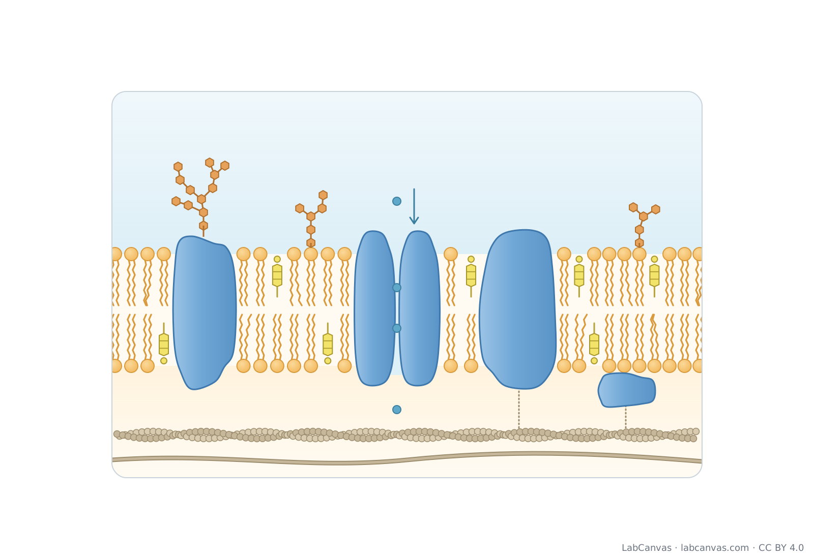
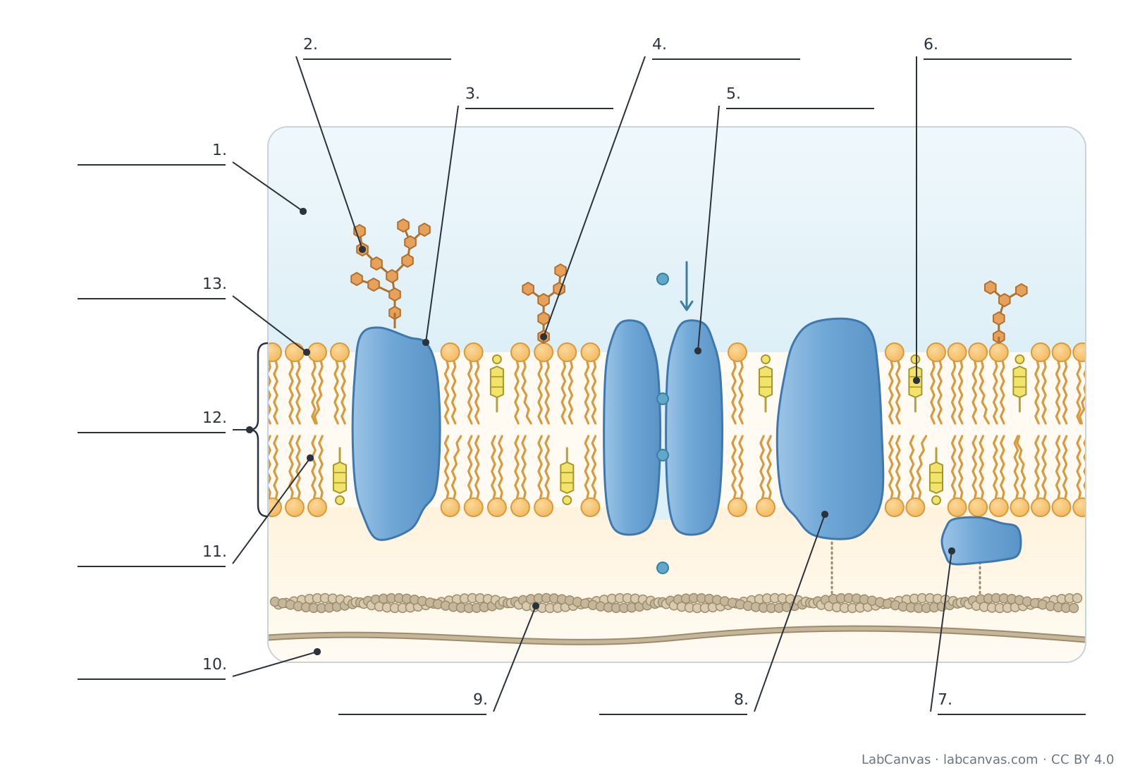
Download the cell membrane diagram
Free to use in homework, teaching, slides, posters and publications under CC BY 4.0. Please credit: Cell membrane diagram by LabCanvas (labcanvas.com), CC BY 4.0
Parts of the cell membrane and what they do
| # | Part | What it is or does |
|---|---|---|
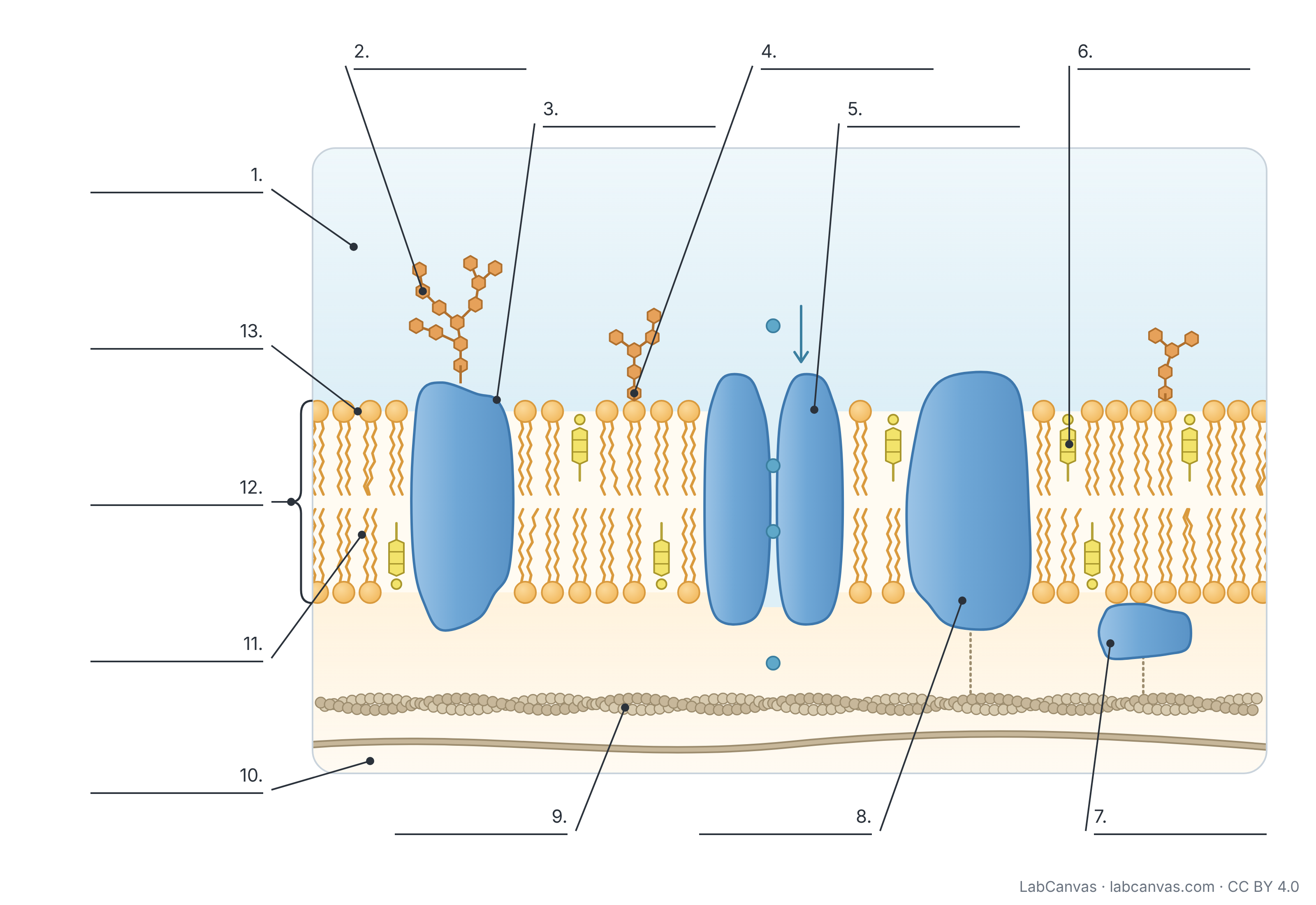
| 1 | Extracellular fluid | The watery fluid outside the cell, on the side of the membrane that carries the carbohydrate chains. |
| 2 | Carbohydrate chain (glycocalyx) | Short, often branched sugar chains on the outer surface; together they form the glycocalyx, which helps cells recognise each other. |
| 3 | Glycoprotein | A membrane protein with carbohydrate chains attached; works in cell recognition and as a receptor. |
| 4 | Glycolipid | A membrane lipid with a carbohydrate chain attached, found only in the outer layer; also helps in cell recognition. |
| 5 | Channel protein | An integral protein with a water-filled pore that lets specific ions or water (aquaporins) cross by facilitated diffusion. |
| 6 | Cholesterol | A lipid wedged between phospholipids that buffers membrane fluidity, keeping it from becoming too fluid or too rigid. |
| 7 | Peripheral protein | A protein on the inner or outer surface, attached to integral proteins or lipids; can act as an enzyme or anchor the cytoskeleton. |
| 8 | Integral (transmembrane) protein | A protein built into the bilayer; transmembrane proteins span it, with hydrophobic middles and hydrophilic ends. |
| 9 | Cytoskeleton filaments | Protein fibres under the membrane that support its shape and anchor some membrane proteins. |
| 10 | Cytoplasm | The inside of the cell, on the side of the membrane facing the cytoskeleton. |
| 11 | Hydrophobic tails | Fatty acid chains that avoid water and face each other in the middle of the bilayer; kinks in unsaturated tails add fluidity. |
| 12 | Phospholipid bilayer | Two layers of phospholipids that form the basic structure of the membrane and a barrier to most polar substances. |
| 13 | Hydrophilic head | The phosphate-containing head of a phospholipid, which faces the watery fluid on each side of the membrane. |
How to label a cell membrane diagram
- Draw the bilayerDraw two parallel rows of small circles (the heads), one near the top and one near the bottom. From each head draw two short wavy lines toward the middle (the tails), so the tails of the two rows meet in the centre.
- Mark the two sidesLabel the top side as extracellular fluid and the bottom side as cytoplasm. Carbohydrate chains always go on the extracellular side.
- Add proteinsDraw large rounded shapes that span the whole bilayer (integral proteins), one with a gap or pore down the middle (channel protein), and a smaller shape stuck to the inner surface only (peripheral protein).
- Add sugars and cholesterolDraw short branched chains of small hexagons on top of one protein (glycoprotein) and on top of one phospholipid head (glycolipid). Add a few small rigid shapes between the tails for cholesterol.
- Add the cytoskeletonDraw a thin beaded filament running under the membrane in the cytoplasm, linked to a membrane protein.
- Label with leader linesPut labels outside the drawing and draw a straight line from each label to its part, with no lines crossing. Use a bracket to label the whole phospholipid bilayer.
Types of membrane proteins
| Type | Where it sits | Main job |
|---|---|---|
| Integral (transmembrane) protein | Built into the bilayer; transmembrane proteins span it from side to side | Transport, signalling (receptors) and links between the inside and outside of the cell |
| Channel protein | An integral protein spanning the bilayer, with a pore through it | Lets specific ions or water cross by facilitated diffusion, with no energy from the cell |
| Peripheral protein | On the inner or outer surface, attached to integral proteins or lipids | Enzymes, attachment points for the cytoskeleton, parts of recognition sites |
| Glycoprotein | A membrane protein with carbohydrate chains on its outer side | Cell recognition and receptors; part of the glycocalyx |
Why it is called the fluid mosaic model
The model was proposed by S. J. Singer and Garth L. Nicolson in 1972. It is fluid because phospholipids and many proteins can drift sideways within their own layer, so the membrane behaves more like a thin film of oil than a rigid sheet. It is a mosaic because proteins, cholesterol and carbohydrate chains are scattered through the bilayer like tiles in a mosaic.
Fluidity matters: the membrane must stay flexible enough for cells such as red and white blood cells to change shape as they squeeze through narrow capillaries. Temperature, the kinks in unsaturated fatty acid tails and cholesterol all affect how fluid it is.
Why phospholipids form a bilayer
A phospholipid has a hydrophilic (water-loving) phosphate head and two hydrophobic (water-fearing) fatty acid tails. In water, phospholipids arrange themselves so that the tails face each other and the heads face out toward the water on both sides. The result is a bilayer whose oily middle blocks ions and most polar molecules, which is why cells need channel and carrier proteins to move these substances across.
What the membrane is made of
The main components are lipids (phospholipids and cholesterol), proteins and carbohydrates attached to some of the lipids and proteins. In a typical human cell, protein makes up about half of the membrane by mass, lipids about 40 percent and carbohydrates the remaining 10 percent (OpenStax Biology 2e, 5.1). The membrane is only about 5 to 10 nm thick, so this diagram is a greatly magnified section.
Quick quiz
1. Which part of a phospholipid faces the watery fluid?
- The hydrophobic tails
- The hydrophilic head
- The cholesterol ring
- The carbohydrate chain
Answer: b) The hydrophilic head. Hydrophilic heads face the extracellular fluid and cytoplasm; the tails face inward.
2. Where are carbohydrate chains found on the cell membrane?
- On the cytoplasm side
- In the middle of the bilayer
- On the extracellular side
- Inside channel proteins
Answer: c) On the extracellular side. Glycoproteins and glycolipids carry their sugar chains on the outer, extracellular surface.
3. What is the main job of a channel protein?
- To make ATP
- To let specific ions or water cross the membrane
- To anchor the cell wall
- To store lipids
Answer: b) To let specific ions or water cross the membrane. Channel proteins have a pore that lets specific substances cross by facilitated diffusion.
4. Which molecule buffers membrane fluidity?
- Cholesterol
- Glucose
- DNA
- Cellulose
Answer: a) Cholesterol. Cholesterol keeps the membrane from becoming too fluid or too rigid.
5. Why is it called the fluid mosaic model?
- The membrane is liquid water
- Its parts can move sideways and proteins are scattered like tiles
- It is made of many cells
- It changes colour
Answer: b) Its parts can move sideways and proteins are scattered like tiles. Fluid: lipids and proteins can move within the layer. Mosaic: many different components scattered through it.
Frequently asked questions
What does a cell membrane diagram show?
It shows a magnified section of the plasma membrane: the phospholipid bilayer with hydrophilic heads and hydrophobic tails, integral, channel and peripheral proteins, cholesterol, and glycoproteins and glycolipids with carbohydrate chains on the outside of the cell.
How do you draw a cell membrane?
Draw two rows of circles with wavy tails pointing inward to make the bilayer, add large protein shapes that span it and a small one on the inner surface, add branched sugar chains on the outer side, add a few cholesterol molecules between the tails, then label the extracellular fluid and cytoplasm.
What are the parts of the cell membrane?
Phospholipids (heads and tails) forming a bilayer, membrane proteins (integral, channel and peripheral), cholesterol, and carbohydrates attached to proteins (glycoproteins) and lipids (glycolipids).
What is the difference between integral and peripheral proteins?
Integral proteins are built into the bilayer, and transmembrane proteins span it completely. Peripheral proteins sit on the inner or outer surface and are attached to integral proteins or lipids rather than passing through the bilayer.
What does cholesterol do in the cell membrane?
Cholesterol sits between phospholipids and acts as a fluidity buffer: it stops the membrane from becoming too fluid when warm and too stiff when cold.
Is the cell membrane the same as the plasma membrane?
Yes. Cell membrane and plasma membrane are two names for the same structure, the membrane that surrounds the cell. All cells have one, including plant cells, where it lies just inside the cell wall.
What is the glycocalyx?
The glycocalyx is the sugar coat on the outside of the cell, made of the carbohydrate chains of glycoproteins and glycolipids. It helps cells recognise each other, for example so the immune system can tell the body’s own cells from foreign ones.
Sources
- OpenStax Biology 2e, 5.1 Components and Structure (CC BY 4.0)
- OpenStax Biology 2e, 5.2 Passive Transport (CC BY 4.0)
- OpenStax Biology 2e, 4.3 Eukaryotic Cells (CC BY 4.0)
Facts on this page follow these introductory textbooks (OpenStax, CC BY 4.0). The diagram is a simplified illustration; organelles are not drawn to scale.
More free diagrams
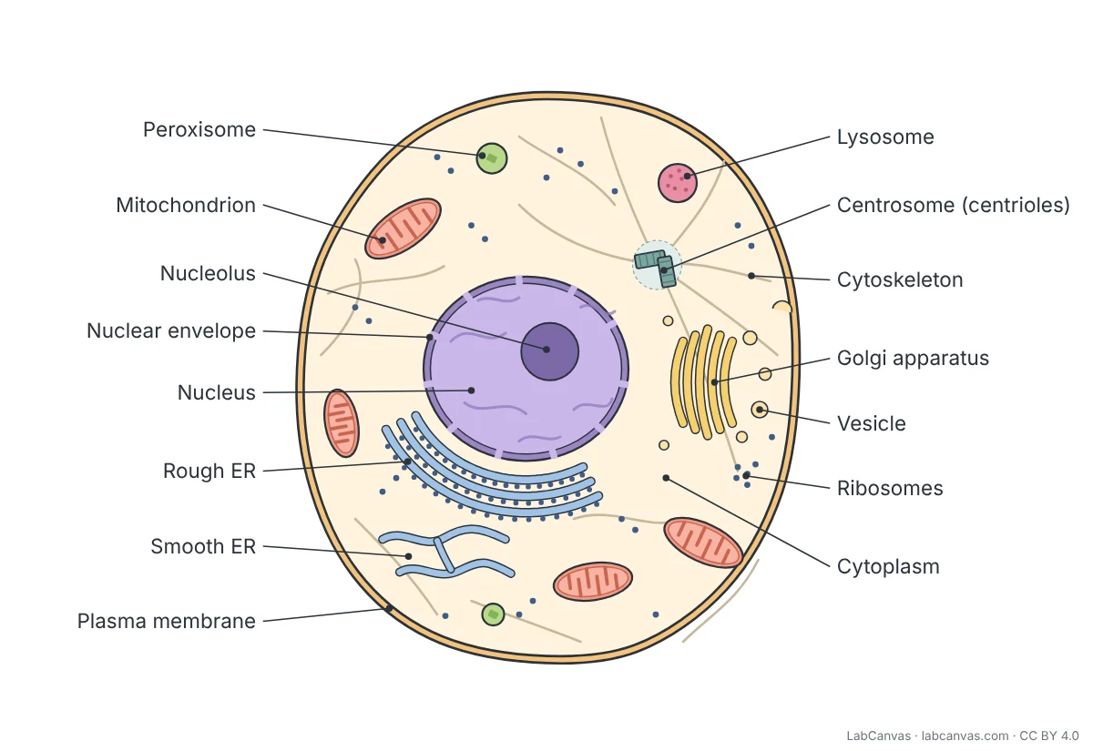
Make your own science diagrams
LabCanvas is a browser workspace for scientific figures, slides and posters, with adjustable cells, organelles and molecules.
Edit this diagram