Meiosis diagram with labels
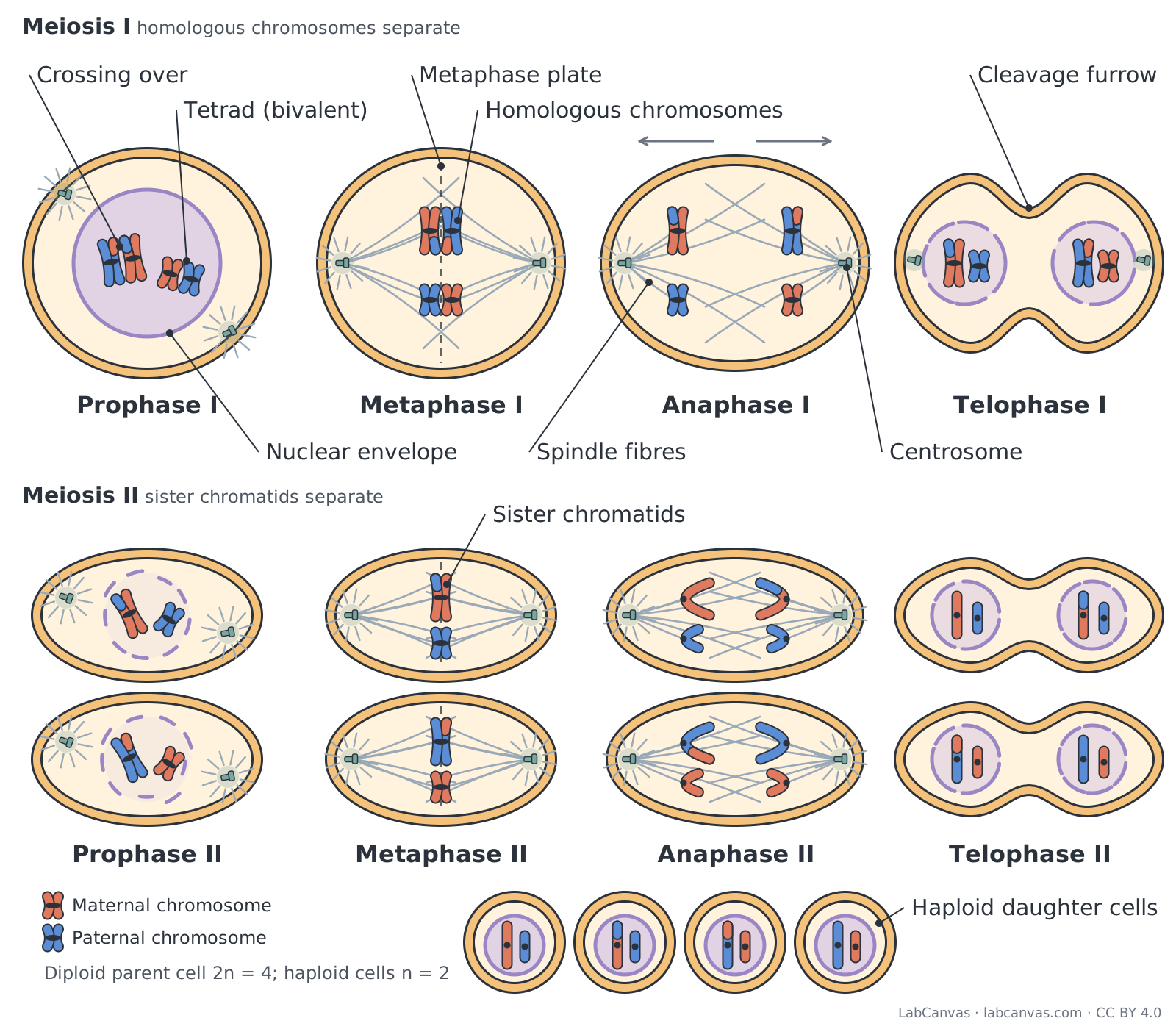
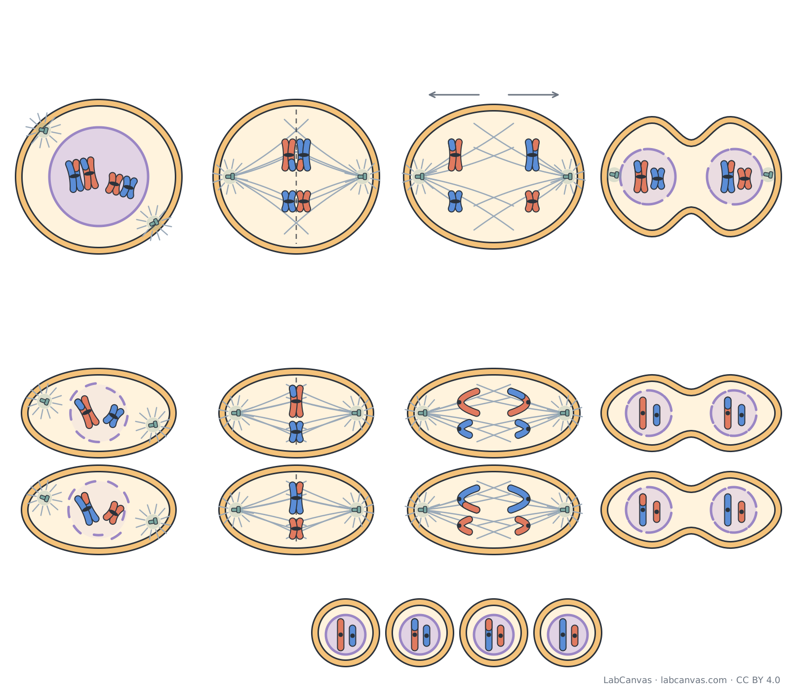
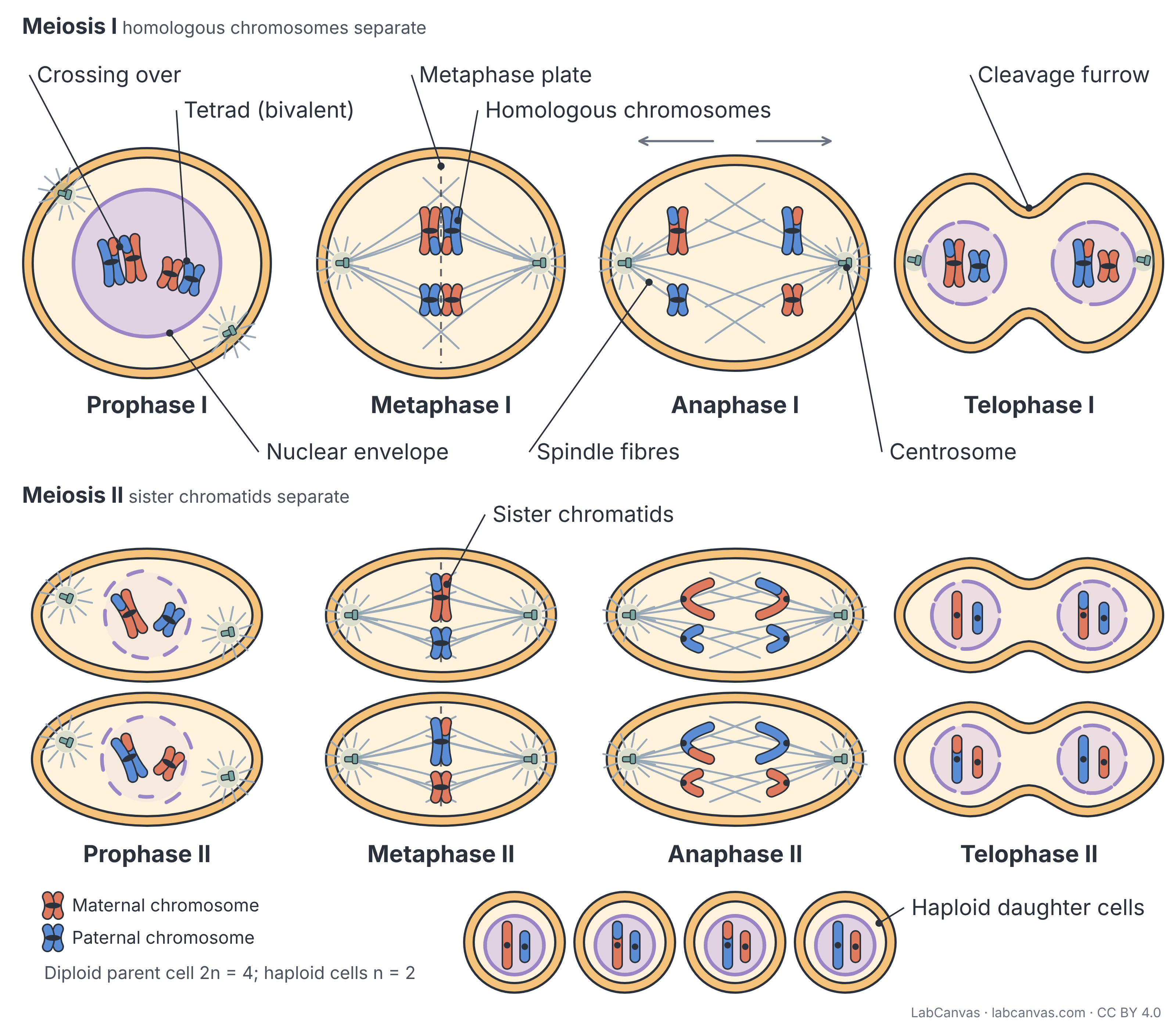
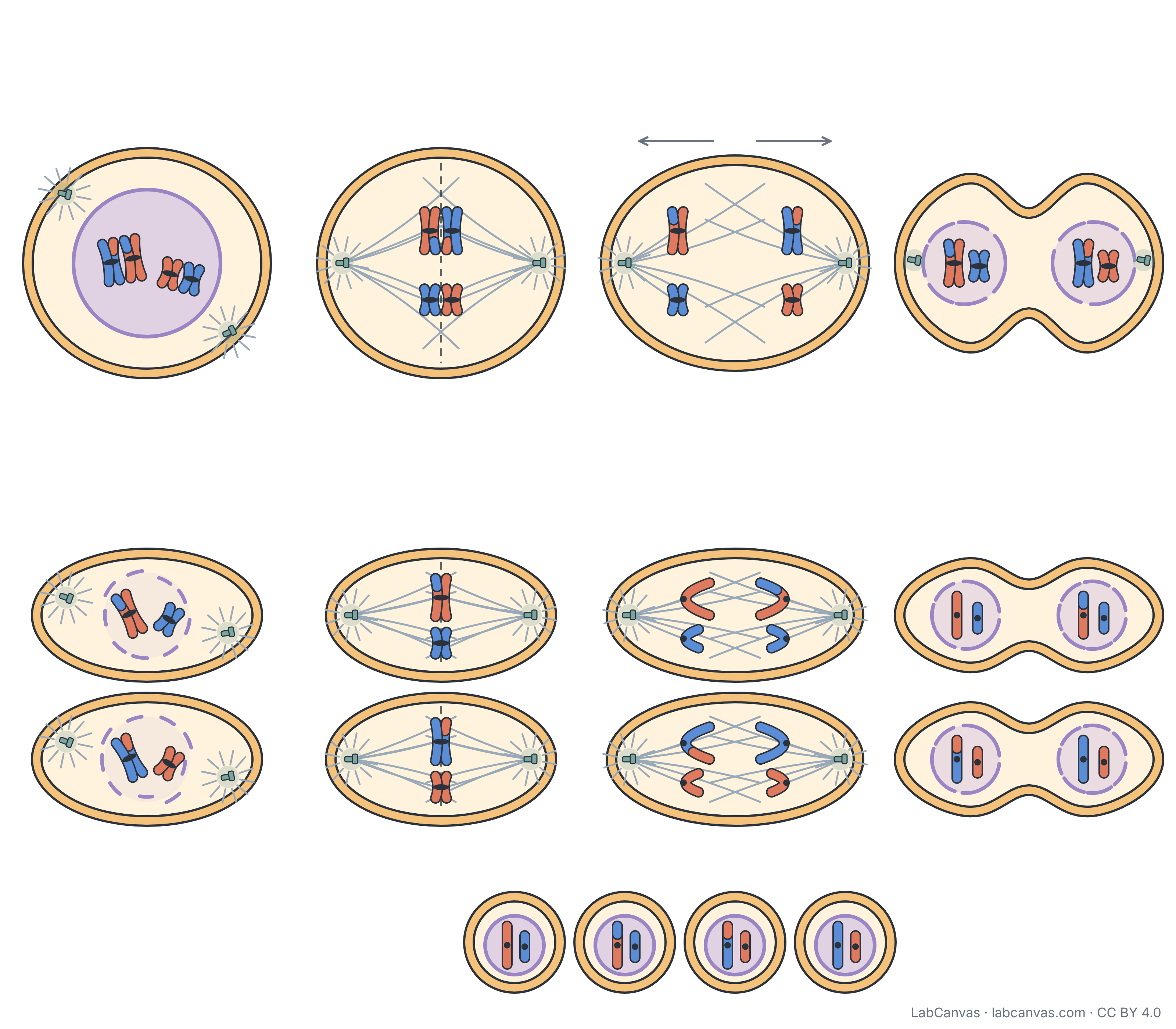
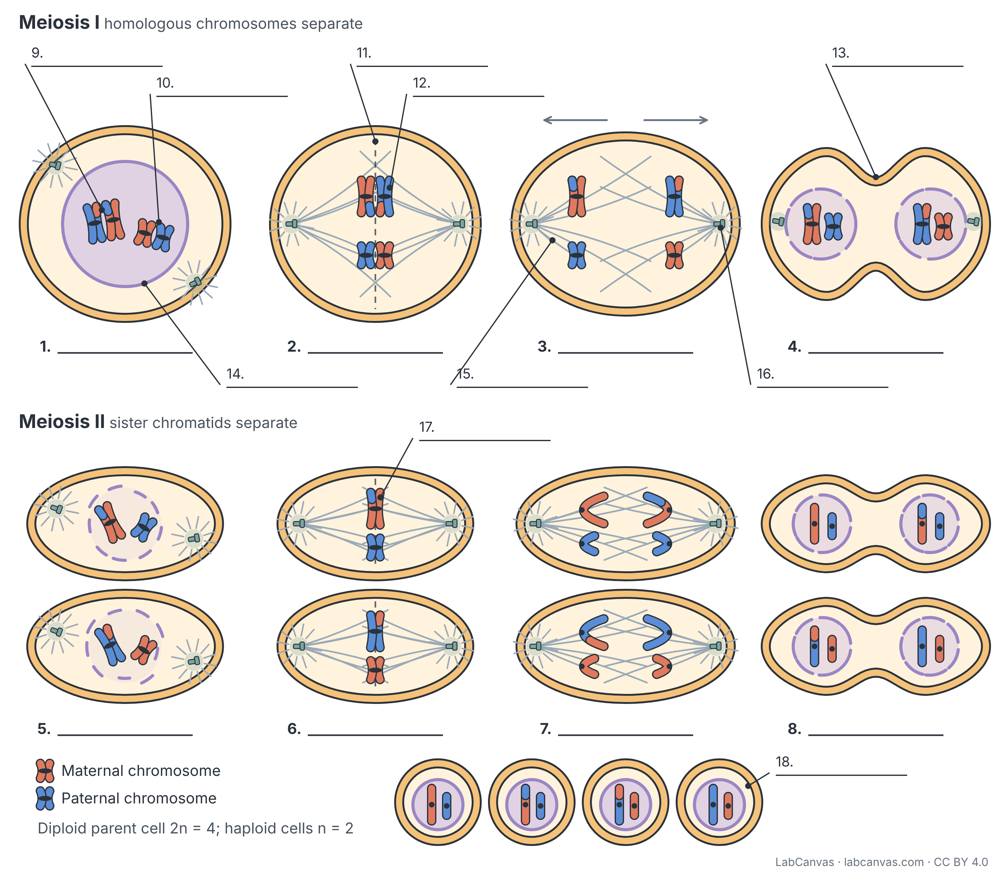
Meiosis is the cell division that turns one diploid cell into four haploid cells, each with half the chromosomes. In this labeled diagram a cell with two homologous pairs (2n = 4) copies its DNA once and then divides twice: meiosis I separates the homologous chromosomes, and meiosis II separates the sister chromatids. Crossing over in prophase I and the random line-up of pairs in metaphase I make all four haploid cells (n = 2) genetically different.
Last updated
Download the meiosis diagram
Free to use in homework, teaching, slides, posters and publications under CC BY 4.0. Please credit: Meiosis diagram by LabCanvas (labcanvas.com), CC BY 4.0
Parts of the meiosis and what they do
| # | Part | What it is or does |
|---|---|---|
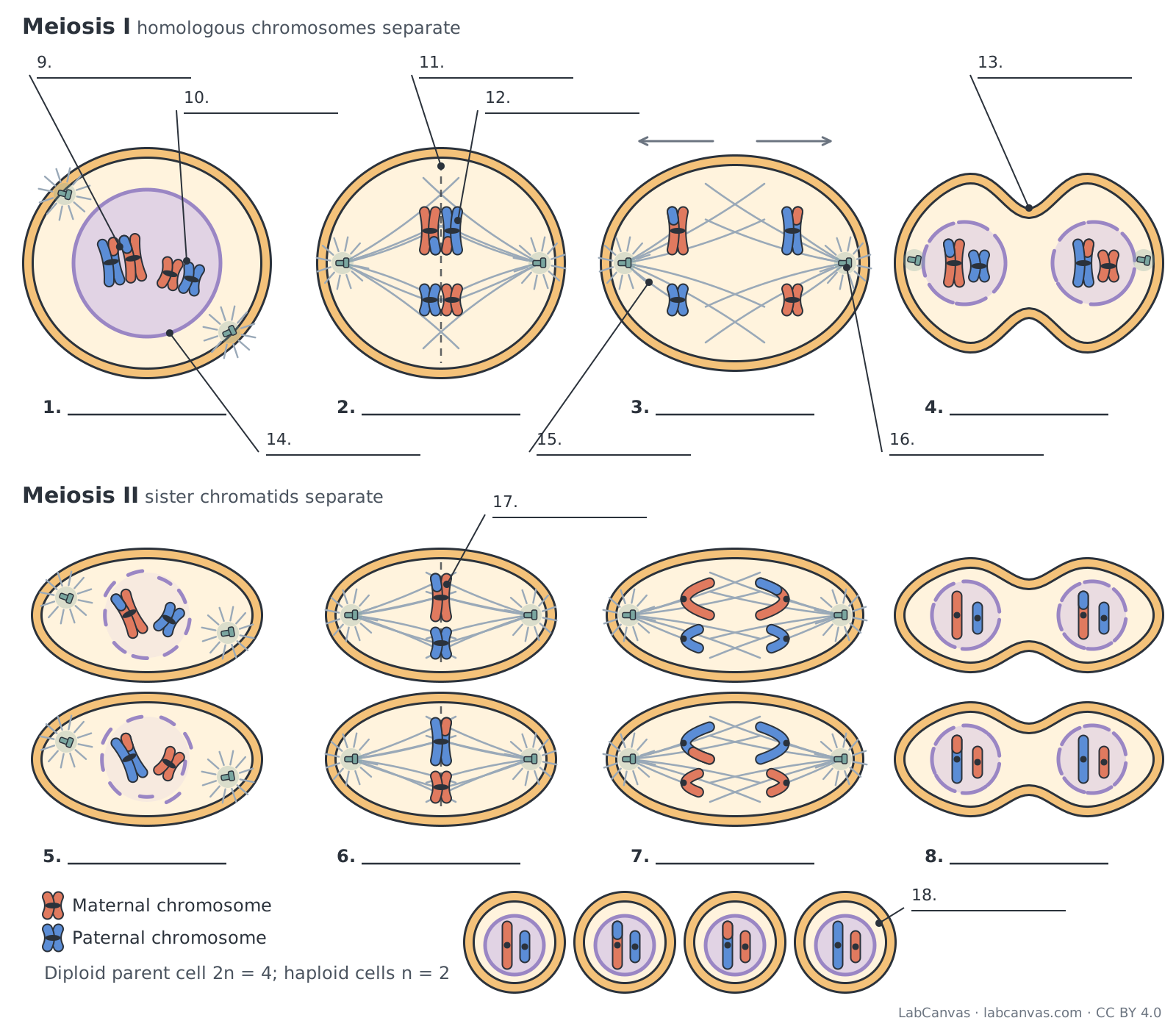
| 1 | Prophase I | Chromosomes condense and each pairs with its homologue (synapsis) to form a tetrad; crossing over swaps segments between non-sister chromatids, and the nuclear envelope starts to break up. |
| 2 | Metaphase I | Homologous pairs line up side by side on the metaphase plate, each homologue facing one pole; the orientation of each pair is random. |
| 3 | Anaphase I | Homologous chromosomes are pulled to opposite poles, while the sister chromatids of each chromosome stay joined at the centromere. |
| 4 | Telophase I | One chromosome of each pair arrives at each pole; in many organisms nuclear envelopes re-form, and cytokinesis gives two haploid cells. |
| 5 | Prophase II | In each haploid cell the chromosomes (still two chromatids each) condense if needed, the nuclear envelope breaks up and a new spindle forms. No DNA is copied before it. |
| 6 | Metaphase II | The chromosomes line up singly on the metaphase plate, with sister chromatids facing opposite poles, as in mitosis. |
| 7 | Anaphase II | The sister chromatids separate and move to opposite poles. |
| 8 | Telophase II | Nuclear envelopes form around the four sets of chromosomes and cytokinesis divides both cells, giving four haploid cells. |
| 9 | Crossing over | Exchange of matching DNA segments between non-sister chromatids of a homologous pair in prophase I; it creates new combinations of alleles. |
| 10 | Tetrad (bivalent) | A pair of synapsed homologous chromosomes in prophase I and metaphase I: two chromosomes, four chromatids. |
| 11 | Metaphase plate | The plane midway between the spindle poles; pairs line up on it in metaphase I and single chromosomes in metaphase II. |
| 12 | Homologous chromosomes | A matching pair of chromosomes, one from each parent, with the same genes in the same order but possibly different alleles. |
| 13 | Cleavage furrow | The groove where the membrane pinches in during cytokinesis of an animal cell, splitting one cell into two. |
| 14 | Nuclear envelope | The double membrane around the nucleus; it breaks up in prophase I and prophase II so the spindle can reach the chromosomes. |
| 15 | Spindle fibres | Microtubules that attach to the chromosomes at their kinetochores and move them; other spindle microtubules overlap between the poles. |
| 16 | Centrosome | The microtubule-organising centre at each spindle pole of an animal cell, containing a pair of centrioles. |
| 17 | Sister chromatids | The two copies of a chromosome made when DNA is copied before meiosis; they stay together through meiosis I and separate in anaphase II. |
| 18 | Haploid daughter cells | The four products of meiosis, each with one chromosome from every homologous pair (n) and a unique mix of maternal and paternal DNA. |
How to label a meiosis diagram
- Set out two rowsDraw meiosis I (prophase I to telophase I) in the top row and meiosis II (prophase II to telophase II) in the bottom row, then four small haploid cells at the end.
- Use the same chromosome set as mitosisStart with four chromosomes: a long and a short pair, each pair with one maternal and one paternal colour. That makes 2n = 4 and n = 2.
- Pair the homologues in prophase IDraw each maternal chromosome lying alongside its paternal partner to make two tetrads, and swap the end segments of the two inner (non-sister) chromatids of one pair to show crossing over.
- Line up pairs, then separate themIn metaphase I put the pairs on the plate side by side; in anaphase I move whole X-shaped chromosomes (not single chromatids) to each pole.
- Repeat the split without copying DNAIn meiosis II each cell lines up its two chromosomes singly and pulls the sister chromatids apart, just as in mitosis.
- Check the counts, then labelEach final cell must have one long and one short chromosome. Label each phase under its cell, then add tetrad, crossing over, homologous chromosomes, sister chromatids and the spindle parts.
Mitosis vs meiosis
| Feature | Mitosis | Meiosis |
|---|---|---|
| Purpose | Growth, repair and asexual reproduction | Making haploid cells (gametes or spores) for sexual reproduction |
| Divisions after one round of DNA copying | One | Two (meiosis I and meiosis II) |
| Cells produced | Two | Four |
| Chromosome number | Same as the parent (diploid stays diploid) | Halved (diploid to haploid) |
| Genetic makeup of the cells | Identical to each other and to the parent | Different from each other and from the parent |
| Homologous chromosomes pair (synapsis) | No | Yes, in prophase I |
| Crossing over | No | Yes, between non-sister chromatids in prophase I |
| Lines up on the metaphase plate | Individual chromosomes | Homologous pairs (metaphase I), then individual chromosomes (metaphase II) |
| Separates in anaphase | Sister chromatids | Homologous chromosomes (anaphase I), then sister chromatids (anaphase II) |
What happens in each phase of meiosis
Before meiosis the cell copies its DNA once, in the S phase of interphase. Meiosis I is the reduction division: in prophase I homologous chromosomes pair up as tetrads and cross over; in metaphase I the pairs line up on the metaphase plate; in anaphase I the homologues separate; and telophase I with cytokinesis gives two haploid cells whose chromosomes still have two chromatids. Meiosis II works like mitosis in each of those cells: prophase II, metaphase II (chromosomes line up singly), anaphase II (sister chromatids separate) and telophase II, ending with four haploid cells.
How to remember the phases (PMAT twice)
Meiosis runs through PMAT (prophase, metaphase, anaphase, telophase) twice: PMAT I, then PMAT II. Some textbooks, including OpenStax Biology 2e, also name a prometaphase I and prometaphase II, when the nuclear envelope has broken up and spindle microtubules attach to the kinetochores; others count this as the end of prophase. A quick check: homologues separate in I, sisters separate in II.
Where the genetic variation comes from
Crossing over in prophase I swaps matching segments between non-sister chromatids, so chromosomes leave meiosis with new mixes of maternal and paternal alleles (the red-and-blue chromosomes in the diagram). Random alignment in metaphase I means each pair lines up with its maternal or paternal member on either side independently of the other pairs; in the diagram the long pair has its maternal chromosome on the left and the short pair has its paternal one on the left. With n pairs there are 2n possible combinations, so 4 for this cell and more than 8 million for the 23 pairs in a human cell, before crossing over is counted.
Meiosis I vs meiosis II
Meiosis I halves the chromosome number because whole homologues, not chromatids, go to each pole: each cell gets one chromosome of every pair. Meiosis II does not change the chromosome number; it separates the two chromatids of each chromosome. There is no S phase between the two divisions.
Quick quiz
1. When does crossing over happen?
- Prophase I
- Metaphase I
- Anaphase II
- Telophase II
Answer: a) Prophase I. Crossing over happens between paired homologues (tetrads) in prophase I.
2. What separates during anaphase I?
- Sister chromatids
- Homologous chromosomes
- Centrioles only
- Nothing; the cell rests
Answer: b) Homologous chromosomes. Homologues go to opposite poles in anaphase I; sister chromatids stay together until anaphase II.
3. A cell with 2n = 4 completes meiosis. How many chromosomes are in each daughter cell?
- 1
- 2
- 4
- 8
Answer: b) 2. Meiosis halves the chromosome number, so each of the four cells has n = 2.
4. What lines up on the metaphase plate in metaphase I?
- Single chromatids
- Homologous pairs (tetrads)
- Single unpaired chromosomes
- Nucleoli
Answer: b) Homologous pairs (tetrads). In metaphase I homologous pairs line up side by side; single chromosomes line up in metaphase II.
5. Which phase of meiosis II looks most like anaphase of mitosis?
- Prophase II
- Metaphase II
- Anaphase II
- Telophase I
Answer: c) Anaphase II. In both, the sister chromatids of each chromosome separate to opposite poles.
Frequently asked questions
What are the stages of meiosis in order?
Meiosis I: prophase I, metaphase I, anaphase I, telophase I. Meiosis II: prophase II, metaphase II, anaphase II, telophase II. Cytokinesis follows each telophase, giving four haploid cells. Some books add prometaphase I and II.
What should be labeled on a meiosis diagram?
Label the eight phases, then homologous chromosomes, tetrad (bivalent), crossing over, sister chromatids, centromere, spindle fibres, centrosome, metaphase plate, nuclear envelope, cleavage furrow and the haploid daughter cells.
How do you show crossing over in a diagram?
Draw the two homologues side by side in prophase I in different colours, then swap the tips of the two inner (non-sister) chromatids so each carries a segment of the other colour. Keep those mixed chromatids through to the final cells.
What is the difference between anaphase I and anaphase II?
In anaphase I homologous chromosomes separate and each still has two sister chromatids. In anaphase II the sister chromatids themselves separate, as in mitosis.
How many chromosomes are in each cell after meiosis?
Half the number in the parent cell. The diagram starts with 4 (2n = 4) and ends with 2 per cell (n = 2); in humans meiosis turns 46 chromosomes into 23 in each egg or sperm.
Why are the four cells made by meiosis different?
Crossing over in prophase I mixes maternal and paternal DNA within chromosomes, and the random orientation of each pair in metaphase I mixes whole chromosomes. Fertilisation adds more variation.
Is this meiosis diagram free to use?
Yes. The labeled, unlabeled and blank worksheet versions are free to download and reuse under CC BY 4.0 with credit to LabCanvas.
Sources
- OpenStax Biology 2e, 11.1 The Process of Meiosis
- OpenStax Biology 2e, 10.2 The Cell Cycle
- OpenStax Biology 2e, Chapter 11 summary
Facts on this page follow these introductory textbooks (OpenStax, CC BY 4.0). The diagram is a simplified illustration; organelles are not drawn to scale.
More free diagrams
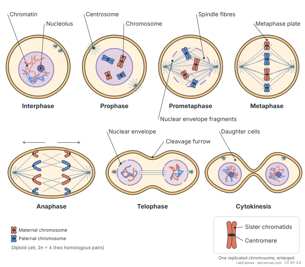
Make your own science diagrams
LabCanvas is a browser workspace for scientific figures, slides and posters, with adjustable cells, organelles and molecules.
Edit this diagram