Mitochondria diagram (labeled structure)
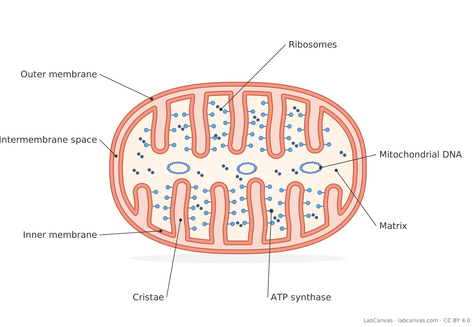
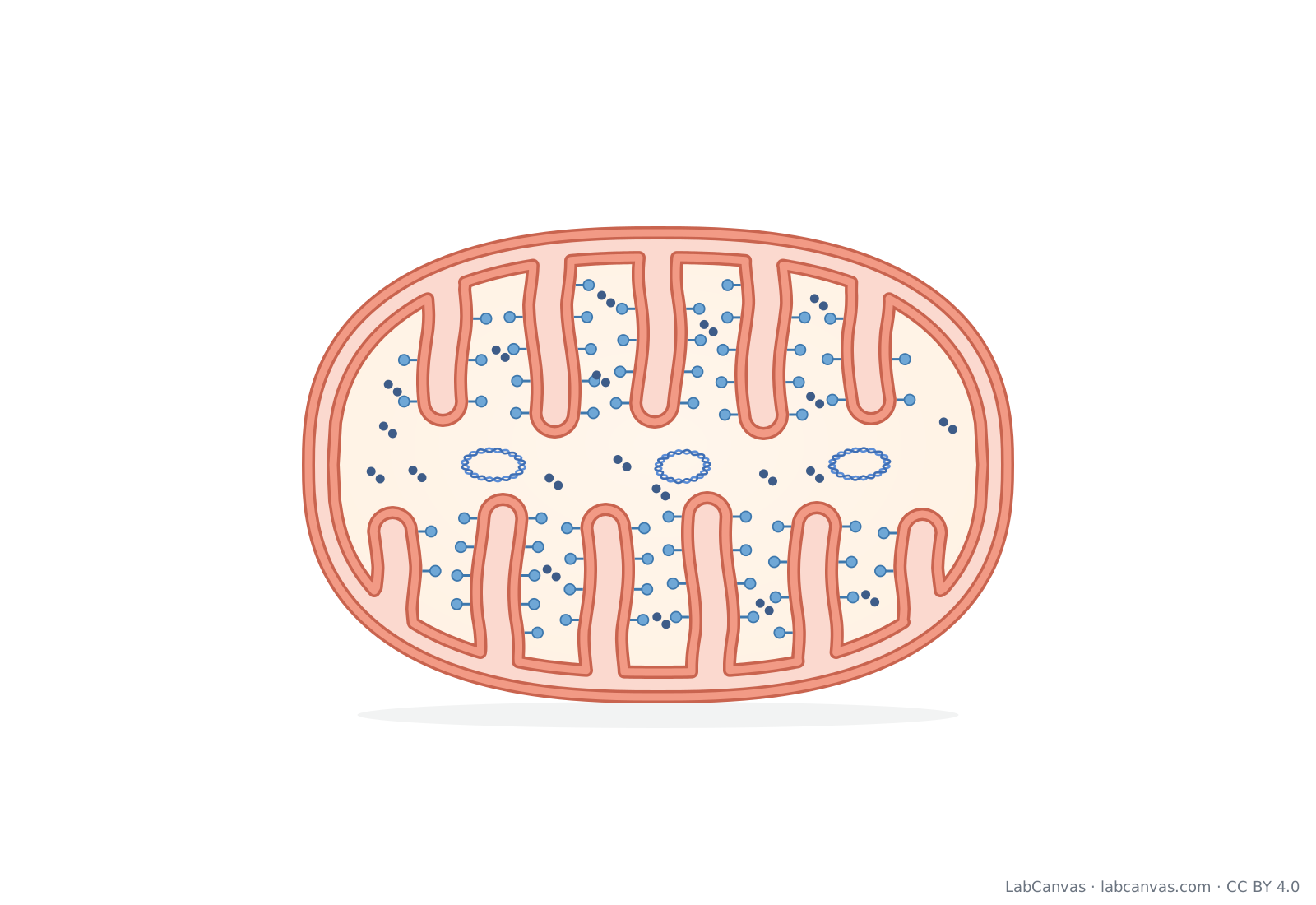
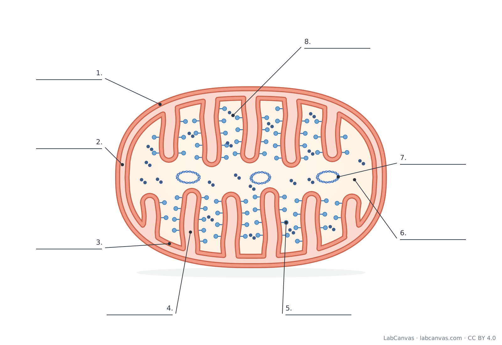
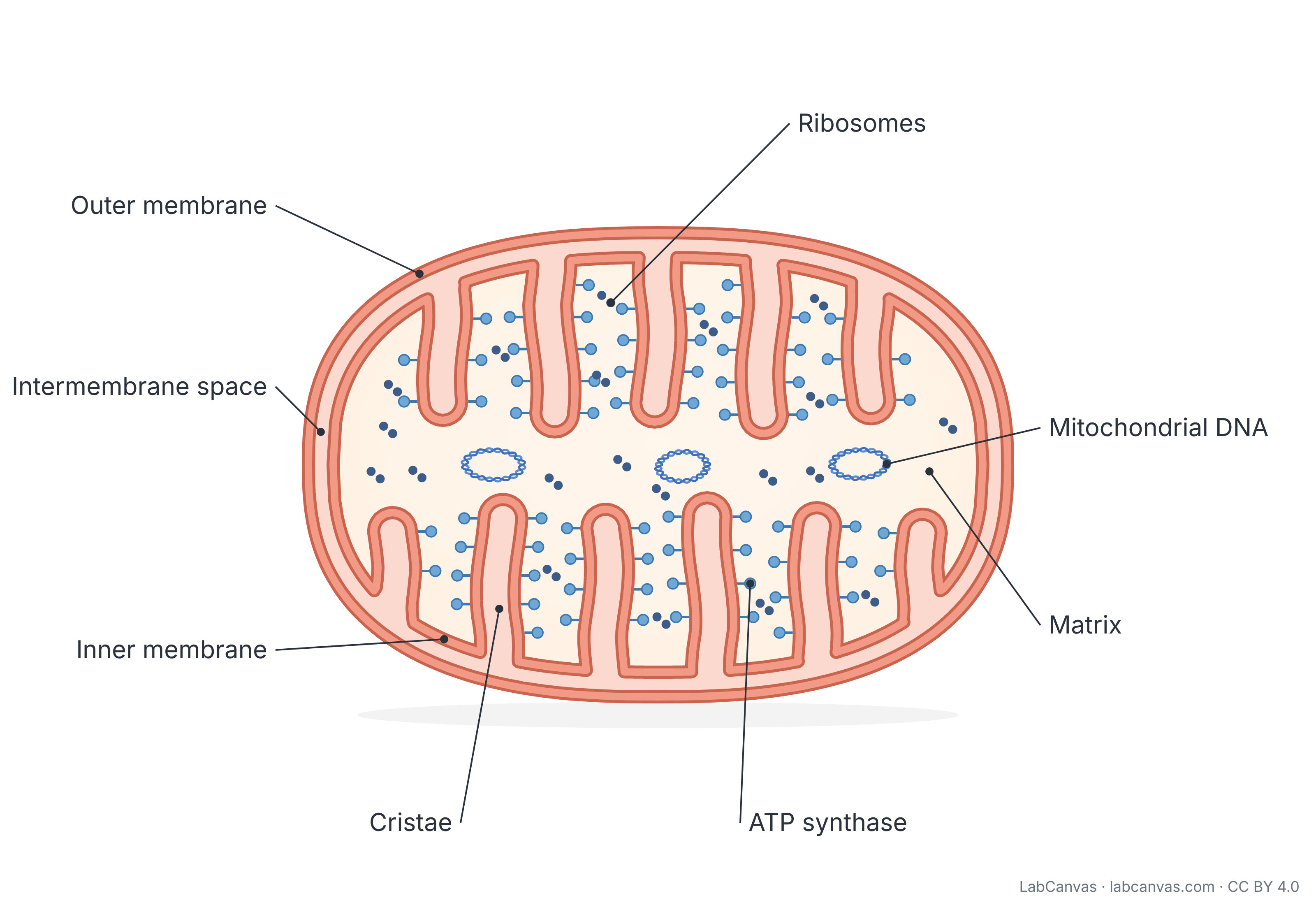
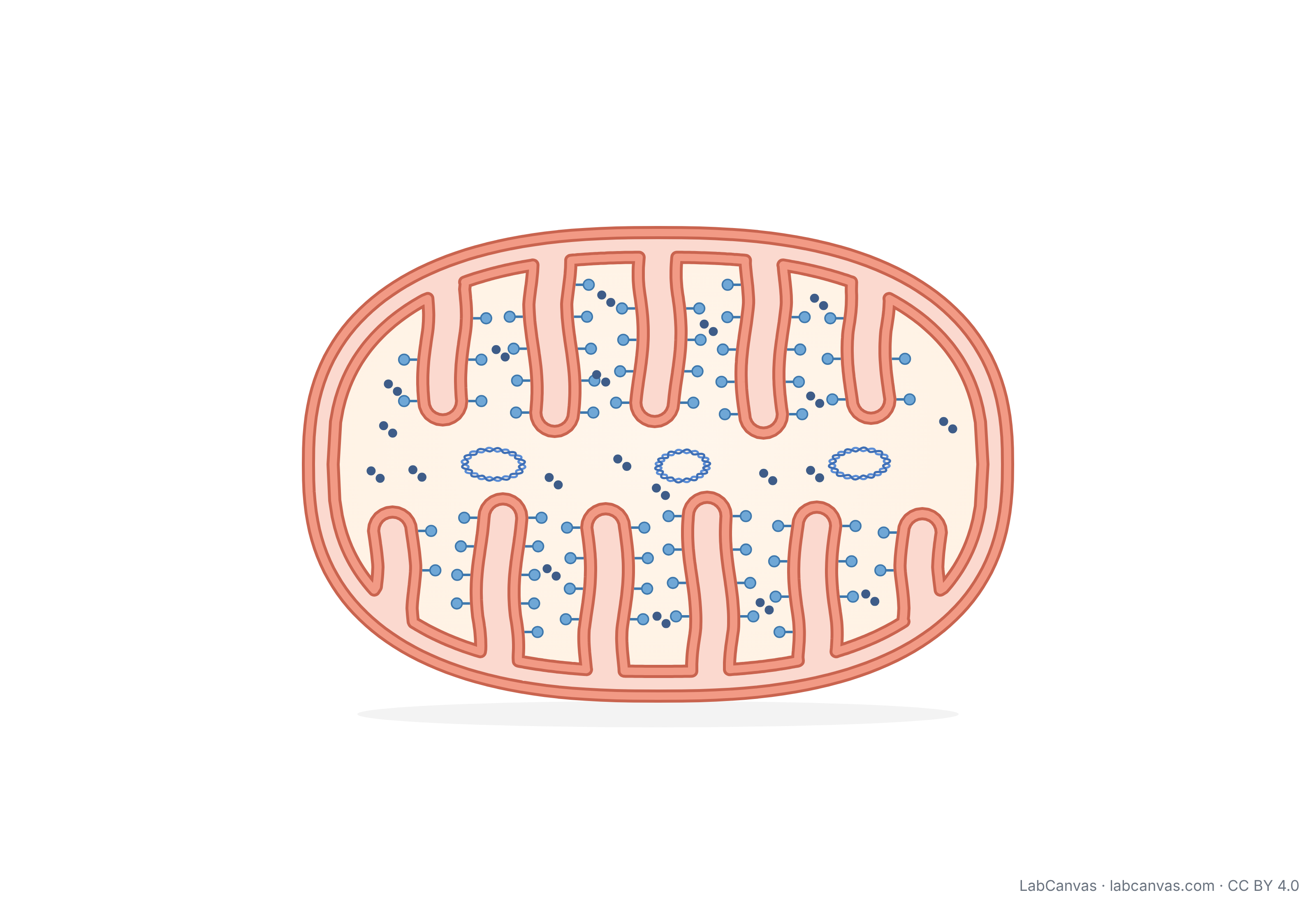
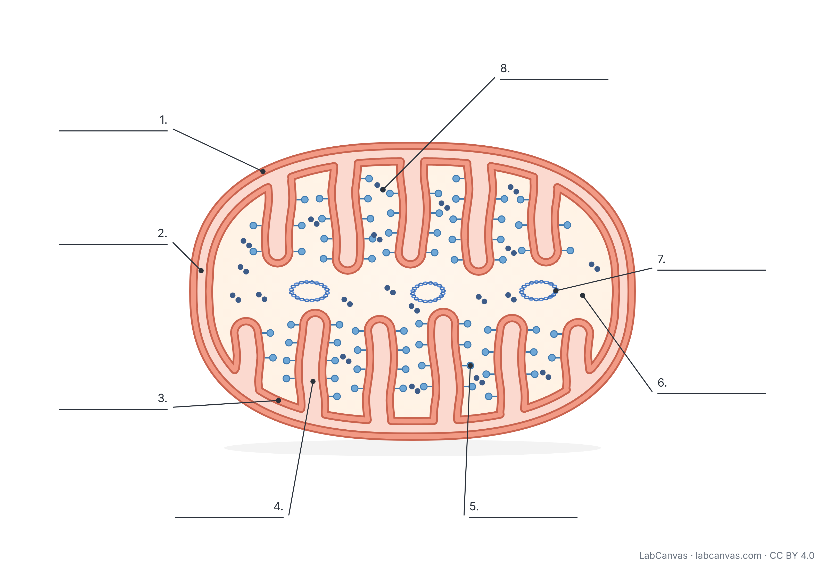
A mitochondria diagram shows a mitochondrion cut lengthways: a smooth outer membrane, a narrow intermembrane space, and an inner membrane folded into cristae that surround the fluid matrix. ATP synthase particles stud the cristae, and the matrix holds the mitochondrion’s own circular DNA and ribosomes. Mitochondria make most of a eukaryotic cell’s ATP by cellular respiration, which is why they are often called the powerhouse of the cell.
Last updated
Download the mitochondria diagram
Free to use in homework, teaching, slides, posters and publications under CC BY 4.0. Please credit: Mitochondria diagram by LabCanvas (labcanvas.com), CC BY 4.0
Parts of the mitochondria and what they do
| # | Part | What it is or does |
|---|---|---|
| 1 | Outer membrane | Smooth phospholipid bilayer that surrounds the whole mitochondrion and separates it from the cytoplasm. |
| 2 | Intermembrane space | The narrow space between the two membranes, where protons (H+) build up during the electron transport chain. |
| 3 | Inner membrane | Second phospholipid bilayer; holds the electron transport chain and ATP synthase and controls what enters the matrix. |
| 4 | Cristae | Folds of the inner membrane that greatly increase its surface area for the electron transport chain and ATP synthase. |
| 5 | ATP synthase | Enzyme complex in the inner membrane that uses the flow of protons back into the matrix to make ATP (chemiosmosis). |
| 6 | Matrix | The fluid inside the inner membrane, where pyruvate is oxidised and the citric acid cycle takes place. |
| 7 | Mitochondrial DNA | The mitochondrion’s own small, circular DNA, which carries genes for some of its own proteins. |
| 8 | Ribosomes | Mitochondrial ribosomes in the matrix that make some of the mitochondrion’s proteins. |
How to label a mitochondria diagram
- Draw the outer membraneDraw a large rounded oval, longer than it is tall. Go over it with a second line just inside to show the membrane as a band.
- Draw the inner membraneDraw a second oval a little inside the first, leaving a narrow gap between them: this gap is the intermembrane space.
- Fold the inner membrane into cristaeInstead of a smooth line, push the inner membrane inward as finger-like folds from the top and bottom edges. Each fold is two lines joined by a rounded tip, and the inside of each fold opens into the intermembrane space.
- Add ATP synthaseDraw small lollipop shapes (a short stalk and a round head) along the cristae, with the heads pointing into the matrix.
- Fill the matrixAdd two or three small closed loops for mitochondrial DNA and small dots for ribosomes in the open space between the cristae.
- Label with leader linesPut the labels around the outside and draw straight lines to each part, with no lines crossing. Make sure the inner membrane label points to the membrane itself, not the space beside it.
Mitochondrion vs chloroplast
| Feature | Mitochondrion | Chloroplast |
|---|---|---|
| Found in | Almost all eukaryotic cells, including plant and animal cells | Plant cells and algae |
| Main process | Cellular respiration: releases energy from food to make ATP | Photosynthesis: captures light energy to make sugars |
| Membranes | Outer and inner membrane | Outer and inner membrane, plus a separate system of thylakoids |
| Inner folds or sacs | Cristae (folds of the inner membrane) | Thylakoids, stacked into grana |
| Fluid inside | Matrix | Stroma |
| Own DNA and ribosomes | Yes | Yes |
| Makes ATP with ATP synthase | Yes, using energy from food molecules | Yes, using light energy |
Stages of cellular respiration and where they happen
Glycolysis happens in the cytoplasm, outside the mitochondrion. It splits one glucose into two pyruvate molecules and gives a small net gain of ATP and NADH.
Pyruvate oxidation and the citric acid cycle happen in the matrix. Pyruvate is carried into the mitochondrion and converted to acetyl CoA, releasing carbon dioxide; the citric acid cycle then releases more carbon dioxide and loads electrons onto NADH and FADH2.
Oxidative phosphorylation happens on the inner membrane and cristae. The electron transport chain passes electrons from NADH and FADH2 to oxygen, the final electron acceptor, which forms water. The energy released pumps protons from the matrix into the intermembrane space. Protons then flow back into the matrix through ATP synthase, which uses that flow to make most of the cell’s ATP. This use of a proton gradient is called chemiosmosis.
Why the inner membrane is folded
The electron transport chain and ATP synthase are built into the inner membrane, so the more inner membrane a mitochondrion has, the more ATP it can make. Folding the membrane into cristae packs a large surface area into a small organelle. Cells that use a lot of energy, such as muscle cells, have many mitochondria.
Why mitochondria have their own DNA
Mitochondria contain their own DNA and ribosomes and divide on their own inside the cell. These features support the endosymbiotic theory: mitochondria are thought to descend from bacteria that were taken up by an ancestral eukaryotic cell and lived inside it. Most mitochondrial proteins are now encoded by genes in the nucleus, but the mitochondrial DNA still carries a small set of its own genes.
Quick quiz
1. What are the folds of the inner mitochondrial membrane called?
- Thylakoids
- Cristae
- Grana
- Villi
Answer: b) Cristae. Cristae are folds of the inner membrane; thylakoids and grana belong to chloroplasts.
2. Where does the citric acid cycle take place?
- In the cytoplasm
- In the intermembrane space
- In the matrix
- In the outer membrane
Answer: c) In the matrix. Pyruvate oxidation and the citric acid cycle happen in the mitochondrial matrix.
3. Where do protons build up during the electron transport chain?
- In the matrix
- In the intermembrane space
- In the nucleus
- Outside the cell
Answer: b) In the intermembrane space. The electron transport chain pumps protons from the matrix into the intermembrane space.
4. What does ATP synthase use to make ATP?
- Light energy directly
- The flow of protons back into the matrix
- Glucose inside the outer membrane
- Ribosomes
Answer: b) The flow of protons back into the matrix. Protons flowing down their gradient through ATP synthase drive ATP production (chemiosmosis).
5. Which feature supports the idea that mitochondria came from bacteria?
- They have cristae
- They have their own DNA and ribosomes
- They are oval
- They are found in muscle cells
Answer: b) They have their own DNA and ribosomes. Their own DNA and ribosomes are key evidence for the endosymbiotic theory.
Frequently asked questions
What does a mitochondria diagram show?
A labelled mitochondria diagram shows the outer membrane, the intermembrane space, the inner membrane folded into cristae, the matrix, ATP synthase particles on the cristae, and the mitochondrial DNA and ribosomes in the matrix.
How do you draw a mitochondrion?
Draw an oval for the outer membrane, a second oval just inside it for the inner membrane, then turn the inner line into finger-like folds (cristae) that reach into the middle. Add small lollipop shapes for ATP synthase, loops for DNA and dots for ribosomes in the matrix, then label each part.
What are the parts of the mitochondria?
Outer membrane, intermembrane space, inner membrane, cristae, matrix, ATP synthase, mitochondrial DNA and ribosomes.
What are cristae and why are they important?
Cristae are the folds of the inner mitochondrial membrane. They increase the surface area available for the electron transport chain and ATP synthase, so the mitochondrion can make more ATP.
What happens in the mitochondrial matrix?
Pyruvate oxidation and the citric acid cycle (Krebs cycle) take place in the matrix. The matrix also contains the mitochondrion’s DNA and ribosomes.
Why are mitochondria called the powerhouse of the cell?
Because they make most of the ATP a eukaryotic cell uses, by breaking down food molecules in cellular respiration. ATP is the energy currency that powers most cell work.
Do plant cells have mitochondria?
Yes. Plant cells have both chloroplasts, for photosynthesis, and mitochondria, for cellular respiration. Almost all eukaryotic cells have mitochondria.
Sources
- OpenStax Biology 2e, 4.3 Eukaryotic Cells (CC BY 4.0)
- OpenStax Biology 2e, 7.2 Glycolysis (CC BY 4.0)
- OpenStax Biology 2e, 7.3 Oxidation of Pyruvate and the Citric Acid Cycle (CC BY 4.0)
- OpenStax Biology 2e, 7.4 Oxidative Phosphorylation (CC BY 4.0)
Facts on this page follow these introductory textbooks (OpenStax, CC BY 4.0). The diagram is a simplified illustration; organelles are not drawn to scale.
More free diagrams
Make your own science diagrams
LabCanvas is a browser workspace for scientific figures, slides and posters, with adjustable cells, organelles and molecules.
Edit this diagram