DNA structure diagram with labels
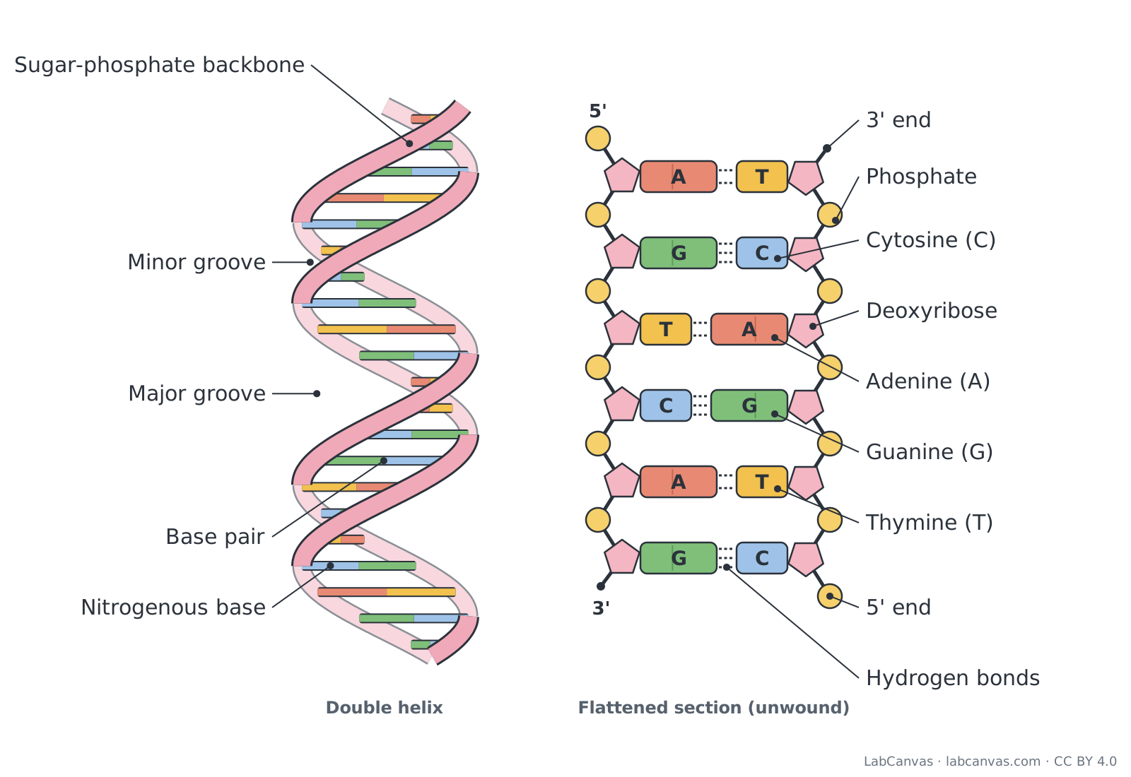
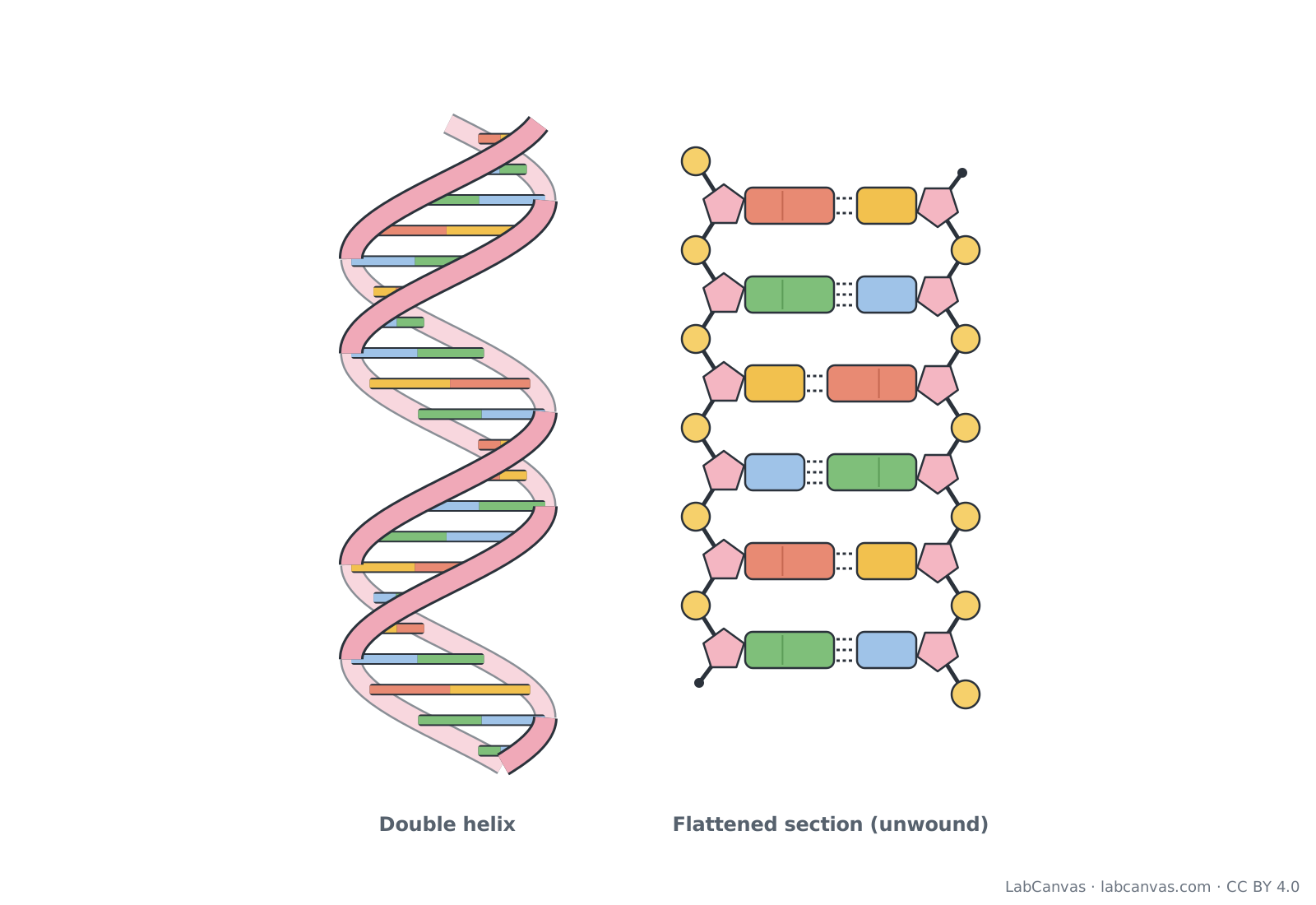
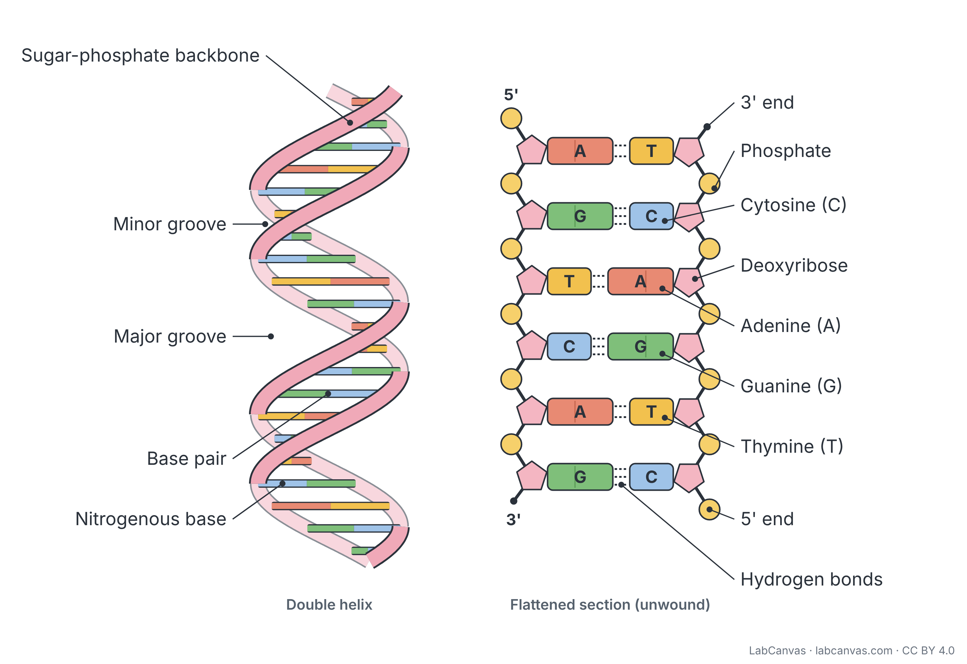
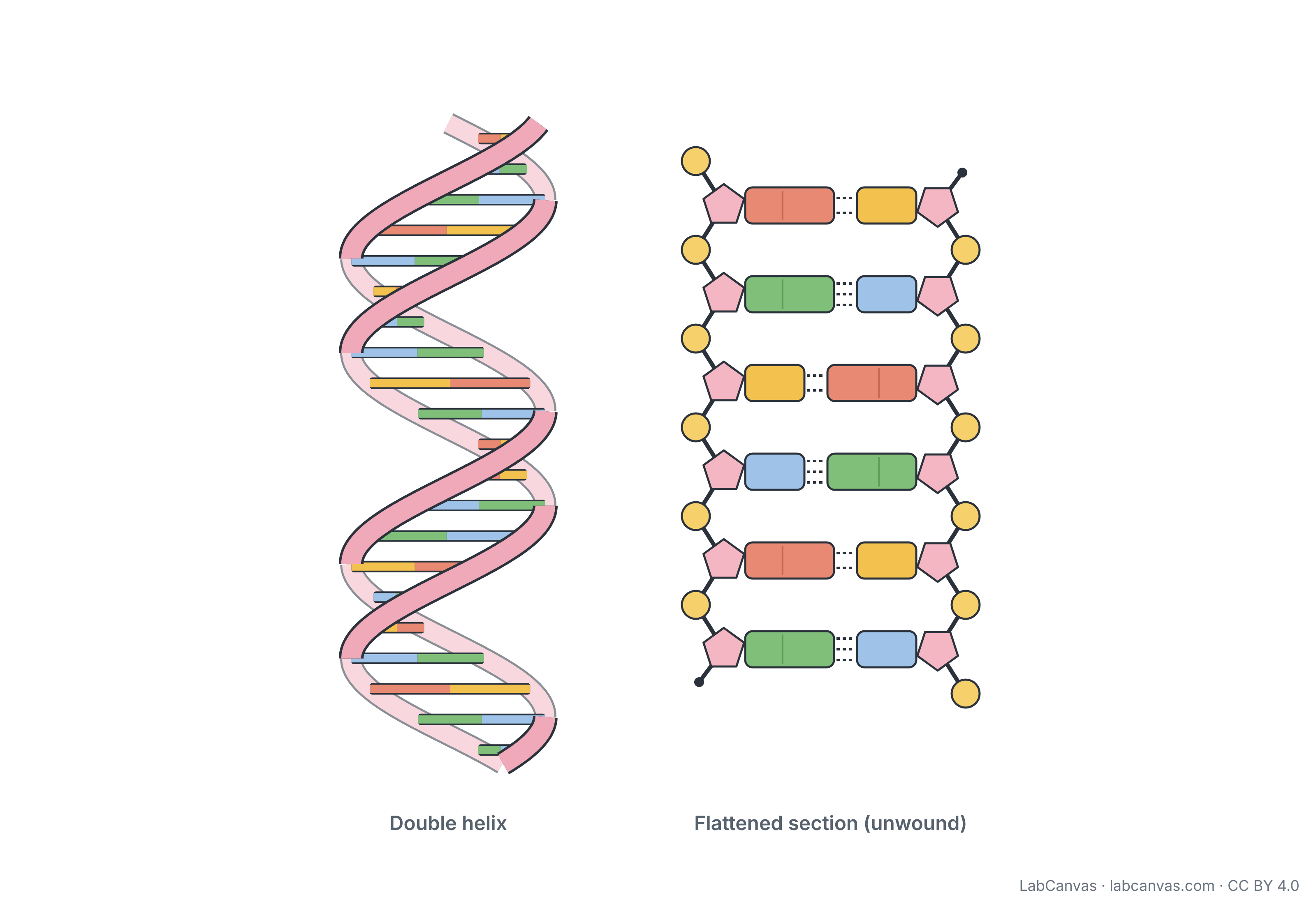
DNA is a double helix: two strands of nucleotides wound around each other. Each strand has a backbone of alternating deoxyribose sugars and phosphates, and the nitrogenous bases point inwards, where they pair by hydrogen bonds, adenine with thymine (2 bonds) and guanine with cytosine (3 bonds). The two strands run in opposite directions (antiparallel), 5' to 3' and 3' to 5', and the twist leaves a major and a minor groove.
Last updated
Download the dna structure diagram
Free to use in homework, teaching, slides, posters and publications under CC BY 4.0. Please credit: DNA structure diagram by LabCanvas (labcanvas.com), CC BY 4.0
Parts of the dna structure and what they do
| # | Part | What it is or does |
|---|---|---|
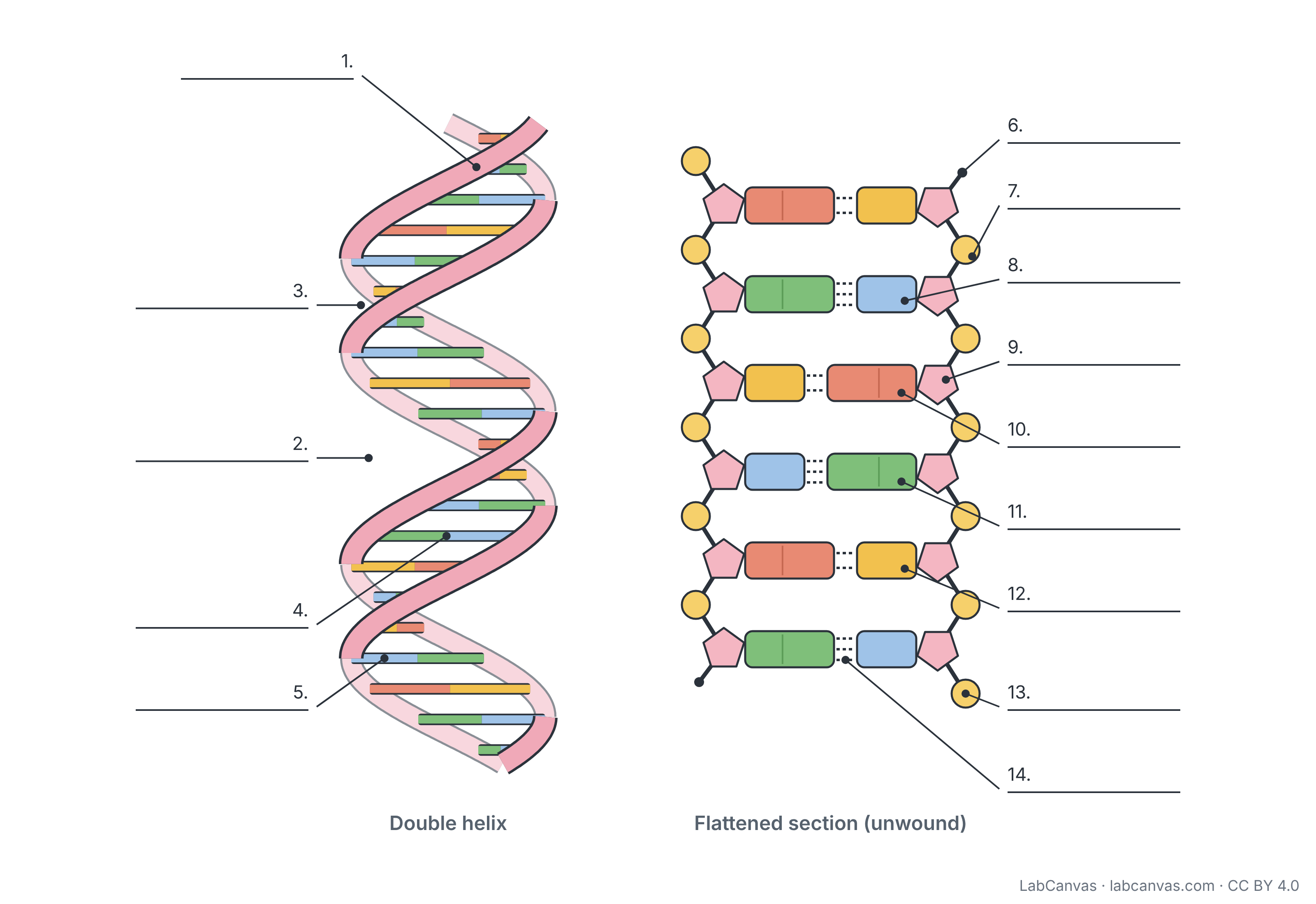
| 1 | Sugar-phosphate backbone | The outside rail of each strand: deoxyribose sugars and phosphates joined in a chain by phosphodiester bonds. |
| 2 | Major groove | The wider of the two grooves that spiral along the helix; proteins that read DNA often bind here. |
| 3 | Minor groove | The narrower groove between the two backbones, alternating with the major groove along the helix. |
| 4 | Base pair | Two complementary bases, one from each strand, joined by hydrogen bonds: A with T or G with C. |
| 5 | Nitrogenous base | The part of a nucleotide that carries information: adenine, thymine, guanine or cytosine; bases point into the helix. |
| 6 | 3' end | The end of a strand with a free -OH on the 3' carbon of the last deoxyribose. |
| 7 | Phosphate | Phosphate group that links the 3' carbon of one sugar to the 5' carbon of the next; it gives DNA its negative charge. |
| 8 | Cytosine (C) | A pyrimidine (single-ring) base; it always pairs with guanine through three hydrogen bonds. |
| 9 | Deoxyribose | The five-carbon sugar of DNA; the base attaches to its 1' carbon and the phosphate to its 5' carbon. |
| 10 | Adenine (A) | A purine (double-ring) base; it always pairs with thymine through two hydrogen bonds. |
| 11 | Guanine (G) | A purine (double-ring) base; it always pairs with cytosine through three hydrogen bonds. |
| 12 | Thymine (T) | A pyrimidine (single-ring) base found in DNA (replaced by uracil in RNA); it pairs with adenine. |
| 13 | 5' end | The end of a strand with a free phosphate on the 5' carbon of the first deoxyribose. |
| 14 | Hydrogen bonds | Weak bonds that hold paired bases together: two between A and T, three between G and C. |
How to label a dna structure diagram
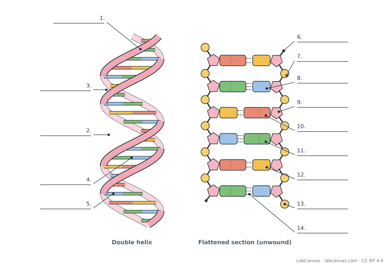
- Start with the railsOn the helix, label one of the twisting ribbons as the sugar-phosphate backbone. On the flattened section, the circles are phosphates and the pentagons are deoxyribose sugars.
- Label the rungsEach rung is a base pair made of two nitrogenous bases. On the ladder, name each base: A pairs with T and G pairs with C, a purine with a pyrimidine.
- Count the hydrogen bondsThe dashed lines between paired bases are hydrogen bonds. Count them to check your pairs: two means A-T, three means G-C.
- Mark the endsFind the strand end with a free phosphate (the 5' end) and the end with a free sugar -OH (the 3' end). Notice that one strand runs 5' to 3' downwards and the other upwards: the strands are antiparallel.
- Find the groovesAlong the edge of the helix the backbones leave gaps of two sizes. Label the wide gap as the major groove and the narrow one as the minor groove, then check the answer key.
A-T vs G-C base pairs
| Feature | A-T pair | G-C pair |
|---|---|---|
| Bases | Adenine and thymine | Guanine and cytosine |
| Purine and pyrimidine | Adenine is the purine, thymine the pyrimidine | Guanine is the purine, cytosine the pyrimidine |
| Hydrogen bonds | 2 | 3 |
| In RNA | Thymine is replaced by uracil, so A pairs with U | Same pairing, G with C |
The building blocks: nucleotides
DNA is a polymer of nucleotides. Each nucleotide has three parts: a five-carbon sugar called deoxyribose, a phosphate group and a nitrogenous base. The carbons of the sugar are numbered 1' to 5': the base is attached to the 1' carbon and the phosphate to the 5' carbon. The bases are of two kinds: the purines adenine and guanine have two rings, and the pyrimidines thymine and cytosine have one. Nucleotides are joined into a strand by phosphodiester bonds between the phosphate of one nucleotide and the 3' carbon of the next sugar.
The double helix
James Watson and Francis Crick proposed the double helix model in 1953, using X-ray diffraction data from Rosalind Franklin and Maurice Wilkins. Two strands twist around each other in a right-handed helix, with the sugar-phosphate backbones on the outside and the bases stacked inside. A purine always pairs with a pyrimidine, so the helix has the same width all along, about 2 nm, and there are about 10 base pairs in each turn. Because the backbones are not evenly spaced around the helix, the twist leaves alternating major and minor grooves.
Complementary and antiparallel strands
Base pairing is specific: A pairs with T and G pairs with C. This makes the two strands complementary, so the sequence of one strand gives the sequence of the other, which is how DNA is copied. The strands are also antiparallel: one runs from its 5' end to its 3' end and the partner runs the opposite way, so the 5' end of one strand sits beside the 3' end of the other.
Quick quiz
1. Which sugar is found in DNA?
- Ribose
- Glucose
- Deoxyribose
- Fructose
Answer: c) Deoxyribose. DNA nucleotides contain the five-carbon sugar deoxyribose.
2. Which base pairs with guanine?
- Adenine
- Thymine
- Uracil
- Cytosine
Answer: d) Cytosine. Guanine pairs with cytosine.
3. How many hydrogen bonds hold an A-T pair together?
- 1
- 2
- 3
- 4
Answer: b) 2. A-T pairs have two hydrogen bonds; G-C pairs have three.
4. What makes up the backbone of a DNA strand?
- Bases and hydrogen bonds
- Sugars and phosphates
- Amino acids
- Bases and sugars only
Answer: b) Sugars and phosphates. Alternating deoxyribose sugars and phosphates form each backbone.
5. The two strands of DNA run in opposite directions. What is this called?
- Complementary
- Semi-conservative
- Antiparallel
- Supercoiled
Answer: c) Antiparallel. Antiparallel strands run 5' to 3' and 3' to 5'.
Frequently asked questions
What are the parts of DNA?
DNA is made of nucleotides, each with a deoxyribose sugar, a phosphate group and a nitrogenous base (A, T, G or C). Sugars and phosphates form the two sugar-phosphate backbones, and the bases pair in the middle by hydrogen bonds, forming base pairs. The twisted molecule has a major and a minor groove and two ends, 5' and 3', on each strand.
What is the structure of DNA?
DNA is a double helix: two antiparallel strands twisted around each other, with sugar-phosphate backbones on the outside and paired bases on the inside, like a twisted ladder.
Which bases pair together in DNA?
Adenine pairs with thymine and guanine pairs with cytosine. Each pair joins a purine (A or G) to a pyrimidine (T or C), which keeps the width of the helix constant.
How many hydrogen bonds are between A and T and between G and C?
Adenine and thymine are joined by two hydrogen bonds; guanine and cytosine are joined by three.
What does antiparallel mean in DNA?
It means the two strands run in opposite directions. One strand runs 5' to 3' and its partner runs 3' to 5', so each end of the double helix has the 5' end of one strand and the 3' end of the other.
What is the difference between the 5' and 3' ends of DNA?
The 5' end has a free phosphate group on the 5' carbon of the sugar; the 3' end has a free hydroxyl (-OH) group on the 3' carbon. New nucleotides are always added to the 3' end.
What are the major and minor grooves of DNA?
They are the two grooves of different widths that spiral along the outside of the double helix between the backbones. Many proteins that bind DNA, such as those that control genes, contact the bases through the grooves.
Sources
- OpenStax Biology 2e, 14.2 DNA Structure and Sequencing (CC BY 4.0)
- OpenStax Biology 2e, 3.5 Nucleic Acids (CC BY 4.0)
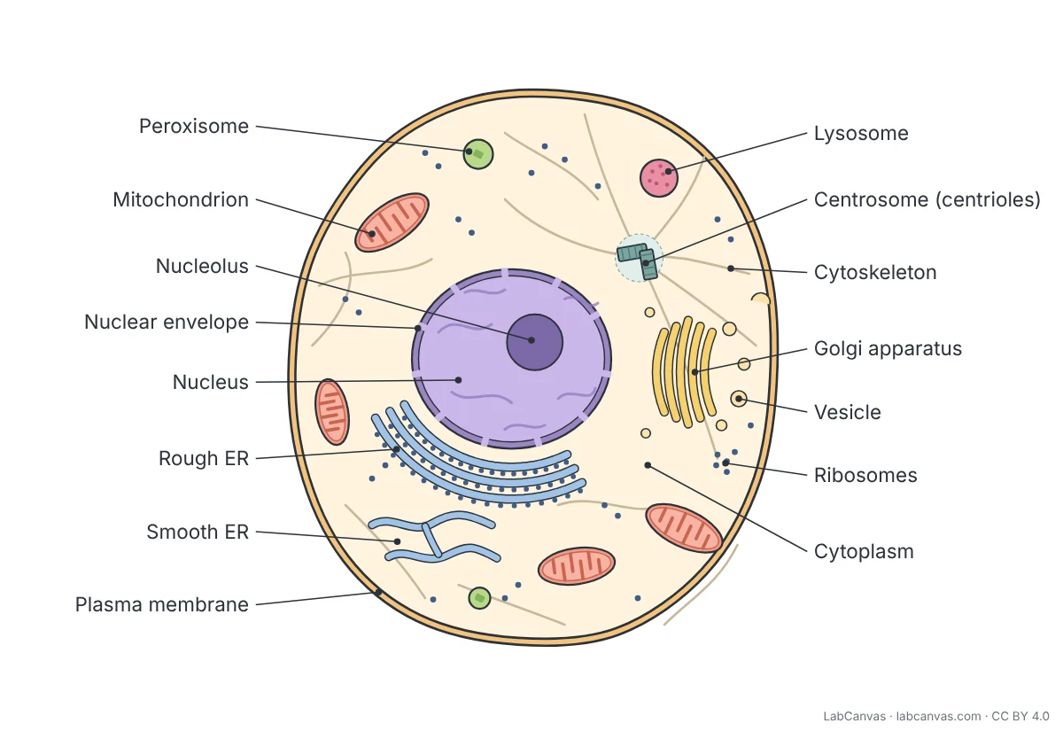
Facts on this page follow these introductory textbooks (OpenStax, CC BY 4.0). The diagram is a simplified illustration; organelles are not drawn to scale.
More free diagrams
Make your own science diagrams
LabCanvas is a browser workspace for scientific figures, slides and posters, with adjustable cells, organelles and molecules.
Edit this diagram