Virus diagram with labels
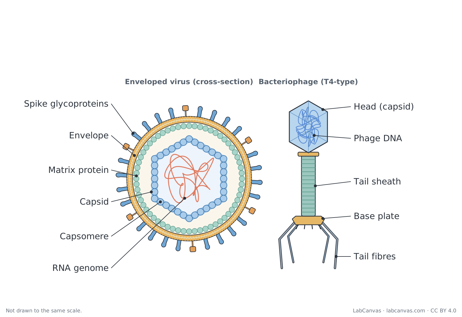
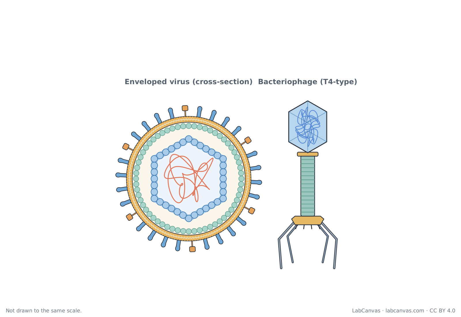
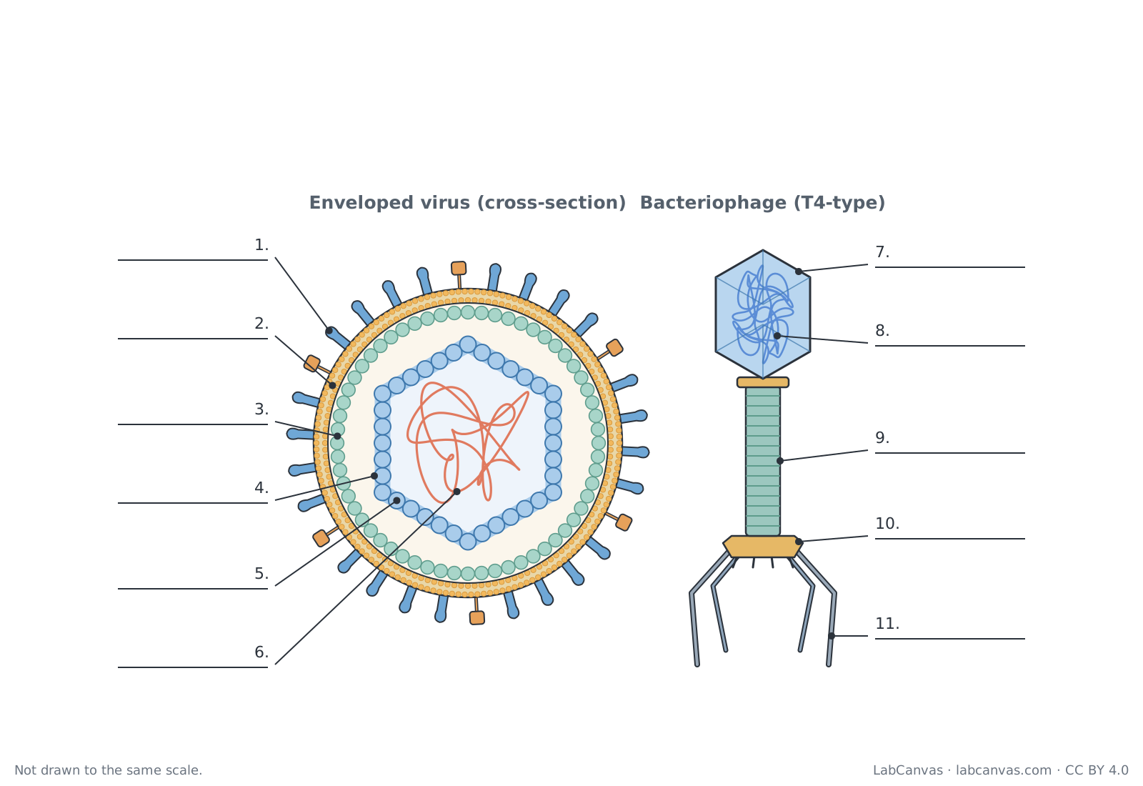
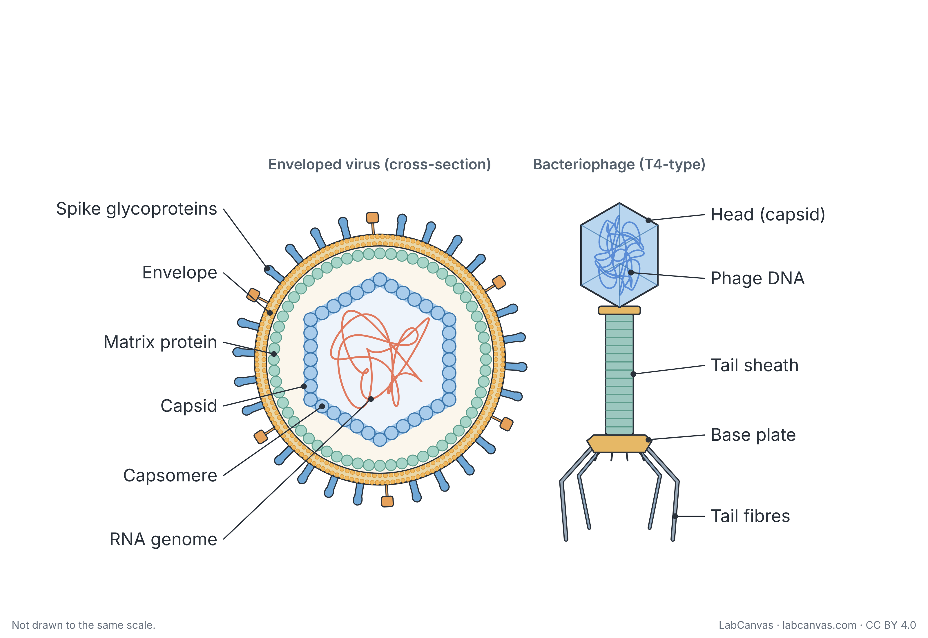
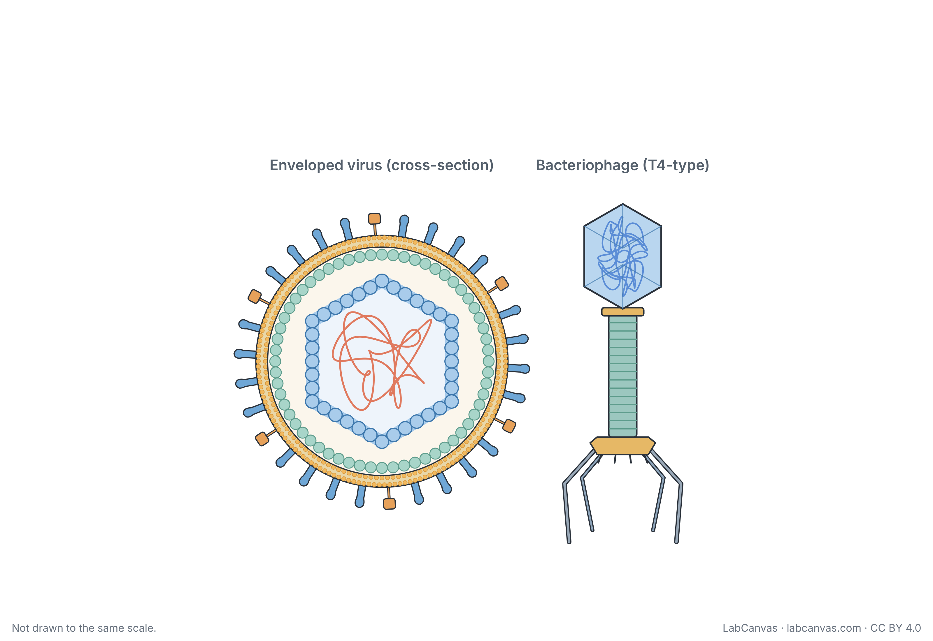
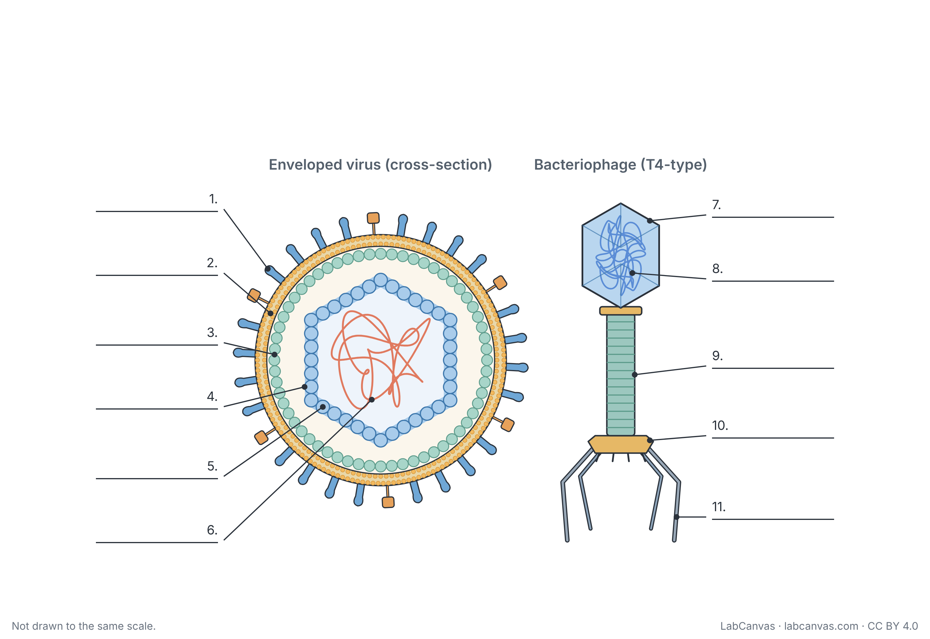
A virus is not a cell: each virus particle (virion) is a genome of DNA or RNA packed inside a protein coat called a capsid, which is built from subunits called capsomeres. Some viruses, such as influenza, are also wrapped in a lipid envelope taken from the host cell and studded with glycoprotein spikes. Bacteriophages, viruses that infect bacteria, often have a head, a tail sheath, a base plate and tail fibres. This free diagram labels both types and comes unlabelled and as a worksheet.
Last updated
Download the virus diagram
Free to use in homework, teaching, slides, posters and publications under CC BY 4.0. Please credit: Virus diagram by LabCanvas (labcanvas.com), CC BY 4.0
Parts of the virus and what they do
| # | Part | What it is or does |
|---|---|---|
| 1 | Spike glycoproteins | Proteins with sugar chains that stick out of the envelope and bind to receptors on a host cell so the virus can attach and enter. |
| 2 | Envelope | Outer lipid bilayer taken from the host cell's membrane as the virus buds out; only some viruses have one. |
| 3 | Matrix protein | Layer of protein just inside the envelope that links the envelope to the capsid and helps the virus assemble and bud. |
| 4 | Capsid | Protein shell that surrounds and protects the viral genome; it is built from many repeated subunits. |
| 5 | Capsomere | One protein subunit of the capsid; many identical capsomeres fit together to form the shell. |
| 6 | RNA genome | The virus's genetic material, here RNA; it carries the instructions the host cell uses to make new virus particles. |
| 7 | Head (capsid) | The bacteriophage's many-sided protein capsid, which holds the phage genome. |
| 8 | Phage DNA | The bacteriophage's genome, packed tightly in the head and injected into the bacterium during infection. |
| 9 | Tail sheath | Hollow protein tube around the tail core; it contracts to drive the core through the bacterial cell wall so the DNA can pass in. |
| 10 | Base plate | Plate at the end of the tail where the tail fibres attach; it anchors the phage to the bacterial surface. |
| 11 | Tail fibres | Long, jointed protein fibres that recognise and bind receptors on the surface of a specific host bacterium. |
How to label a virus diagram
- Start with the outside of the enveloped virusLabel the knobs sticking out as spike glycoproteins and the membrane they sit in as the envelope.
- Move inwardsMark the layer of beads lining the envelope as matrix protein. The many-sided shell inside it is the capsid, and each bead of that shell is a capsomere.
- Find the genomeThe coiled strand inside the capsid is the RNA genome.
- Label the bacteriophage from top to bottomThe many-sided head is the capsid and the coiled strand in it is the phage DNA. Below the head, the ringed tube is the tail sheath, the plate at its end is the base plate and the long jointed legs are the tail fibres.
- Check the two panels separatelyBoth viruses have a protein capsid around a nucleic acid genome; only the enveloped virus has an envelope, and only the phage has a tail. Check your answers against the key.
Virus vs bacterium
| Feature | Virus | Bacterium |
|---|---|---|
| Made of cells? | No; a virion is a genome in a protein capsid, sometimes with an envelope | Yes; a single prokaryotic cell |
| Genetic material | DNA or RNA, never both | DNA (and RNA made from it) |
| Ribosomes and metabolism | None; uses the host cell's ribosomes, enzymes and energy | Own 70S ribosomes and metabolism |
| Reproduction | Only inside a host cell | Divides on its own by binary fission |
| Size | Much smaller; most virions are about 20 to 250 nanometres across | Usually about 0.1 to 5.0 micrometres |
| Treated with antibiotics? | No | Often, yes |
The basic structure of a virus
Every virion has a genome of DNA or RNA, never both, enclosed in a protein coat called the capsid. The capsid is built from many copies of protein subunits called capsomeres. Capsids come in a few basic shapes: helical (rod-like), polyhedral (often icosahedral, with 20 triangular faces) and complex shapes such as the head-and-tail bacteriophages.
Some viruses are enveloped: as they leave the host cell they wrap themselves in a piece of its membrane, which carries viral glycoproteins. These spikes bind to receptors on the next host cell, and a matrix protein layer links the envelope to the capsid. Viruses without an envelope are called naked viruses.
Influenza as an example
Influenza is an enveloped RNA virus. Its envelope carries two kinds of spike glycoprotein, hemagglutinin and neuraminidase, which are drawn here as two spike shapes. Under the envelope is a layer of matrix protein. The diagram shows the capsid as a simple shell for clarity; in influenza the RNA genome is in several separate segments, each wrapped in protein as a helical nucleocapsid.
How a bacteriophage infects a bacterium
A bacteriophage (phage) is a virus that infects bacteria. A T4-type phage attaches to a host bacterium with its tail fibres, which bind receptors on the cell surface, and settles on its base plate. The tail sheath then contracts, pushing the hollow tail core through the cell wall, and the DNA in the head passes into the cell. The empty capsid stays outside while the host cell is made to produce new phages.
Quick quiz
1. What is the protein coat around a viral genome called?
- Envelope
- Capsid
- Cell wall
- Matrix
Answer: b) Capsid. The capsid is the protein shell that encloses the viral DNA or RNA.
2. Where does a virus envelope come from?
- It is made of peptidoglycan
- It is taken from the host cell's membrane
- It is made by the virus's own ribosomes
- It is part of the capsid
Answer: b) It is taken from the host cell's membrane. Enveloped viruses gain their lipid envelope from a host cell membrane as they bud out.
3. What do the spike glycoproteins of an enveloped virus do?
- Store the genome
- Make ATP
- Bind receptors on a host cell
- Digest the cell wall
Answer: c) Bind receptors on a host cell. Glycoprotein spikes attach the virus to specific receptors on its host cell.
4. Which part of a bacteriophage contracts to inject DNA into a bacterium?
- Tail fibres
- Head
- Base plate
- Tail sheath
Answer: d) Tail sheath. The tail sheath contracts, driving the tail core through the cell wall so the DNA can enter.
5. What are the protein subunits of a capsid called?
- Capsomeres
- Ribosomes
- Plasmids
- Nucleotides
Answer: a) Capsomeres. A capsid is assembled from many capsomeres.
Frequently asked questions
What are the parts of a virus?
All viruses have a genome of DNA or RNA inside a protein capsid made of capsomeres. Enveloped viruses also have a lipid envelope with glycoprotein spikes, often with a matrix protein layer beneath it. Bacteriophages such as T4 have a head, a tail sheath, a base plate and tail fibres.
Is a virus a living cell?
No. A virus is not made of cells and has no ribosomes or metabolism of its own. It can only make copies of itself inside a living host cell, using that cell's machinery.
What is the capsid of a virus?
The capsid is the protein coat that surrounds and protects the viral genome. It is made of many protein subunits called capsomeres, and its shape can be helical, polyhedral or complex.
What is the difference between an enveloped and a naked virus?
An enveloped virus is surrounded by a lipid envelope taken from the host cell's membrane, with viral glycoproteins in it. A naked (non-enveloped) virus has only its protein capsid on the outside.
What is a bacteriophage?
A bacteriophage, or phage, is a virus that infects bacteria. Many, such as T4, have a many-sided head holding DNA, a tail with a contractile sheath, a base plate and tail fibres that attach to the host.
Do viruses have DNA or RNA?
A virus has either DNA or RNA as its genome, never both. Influenza and HIV have RNA genomes, while T4 bacteriophage and herpesviruses have DNA genomes.
Why don't antibiotics work on viruses?
Antibiotics target structures and processes of bacteria, such as the peptidoglycan cell wall and 70S ribosomes. Viruses have none of these; they rely on the host cell's machinery, so antibiotics do not affect them.
Sources
- OpenStax Biology 2e, 21.1 Viral Evolution, Morphology, and Classification (CC BY 4.0)
- OpenStax Microbiology, 6.1 Viruses (CC BY 4.0)
- OpenStax Biology 2e, 21.2 Virus Infections and Hosts (CC BY 4.0)
- OpenStax Biology 2e, 4.2 Prokaryotic Cells (CC BY 4.0)
Facts on this page follow these introductory textbooks (OpenStax, CC BY 4.0). The diagram is a simplified illustration; organelles are not drawn to scale.
More free diagrams
Make your own science diagrams
LabCanvas is a browser workspace for scientific figures, slides and posters, with adjustable cells, organelles and molecules.
Edit this diagram