Heart diagram with labels
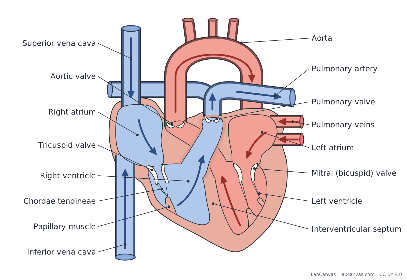
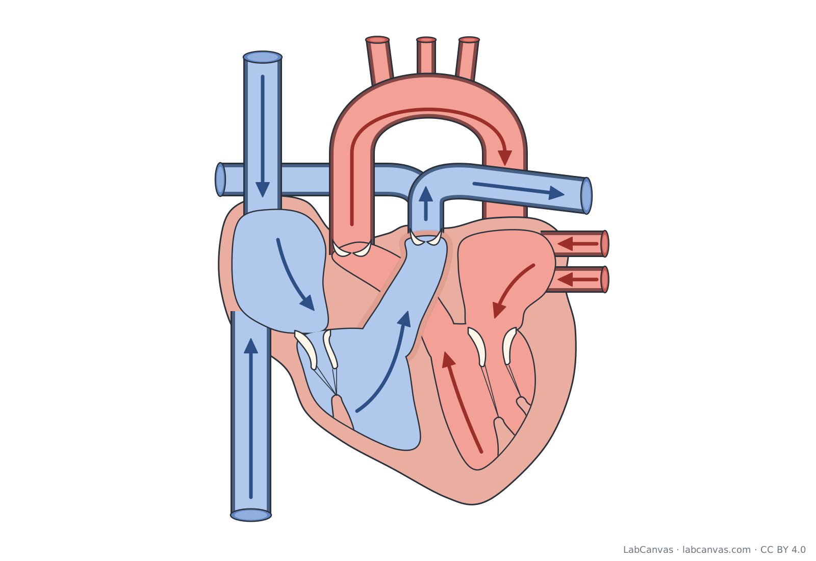
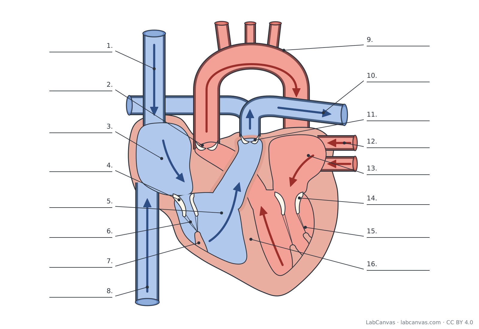
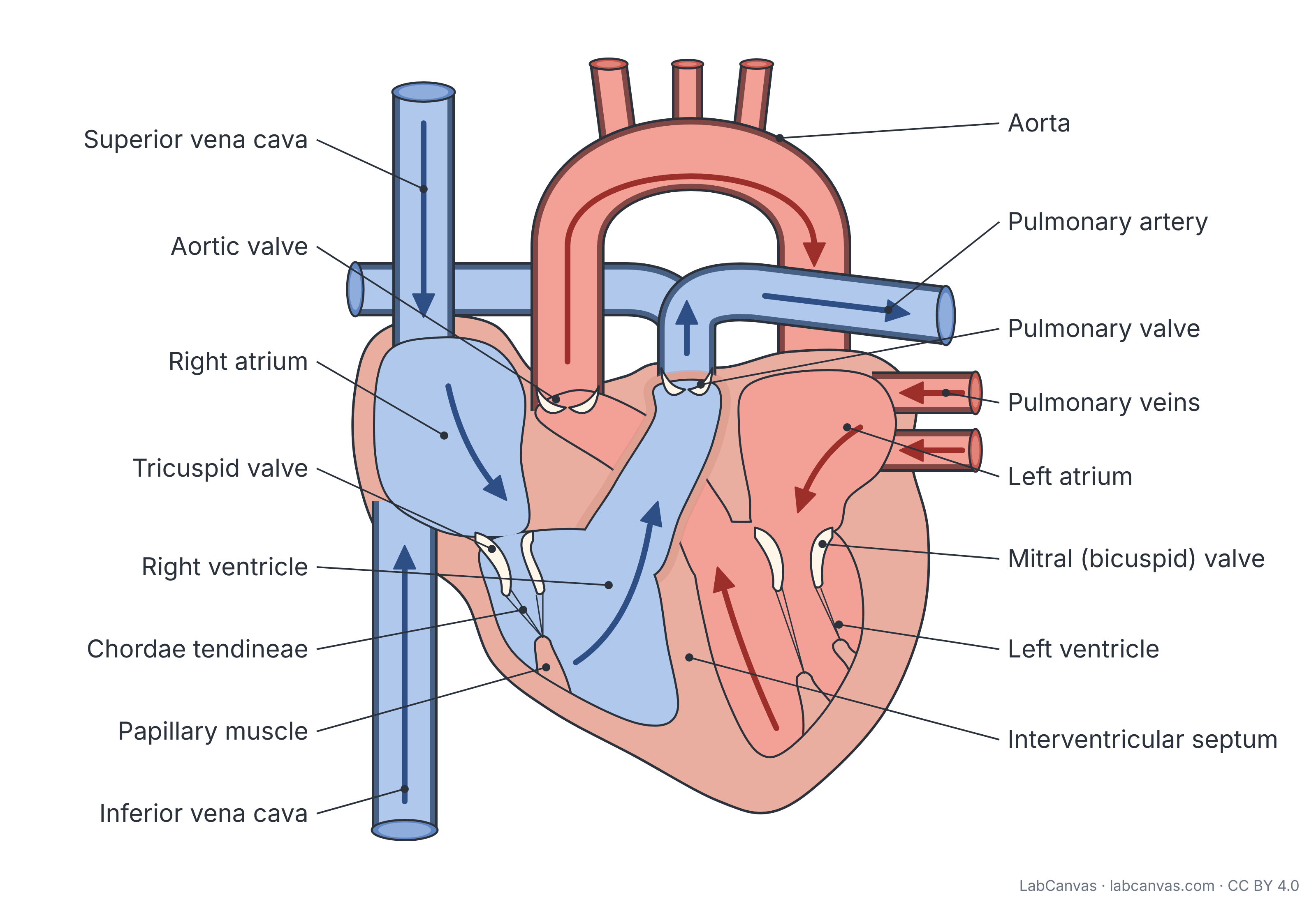
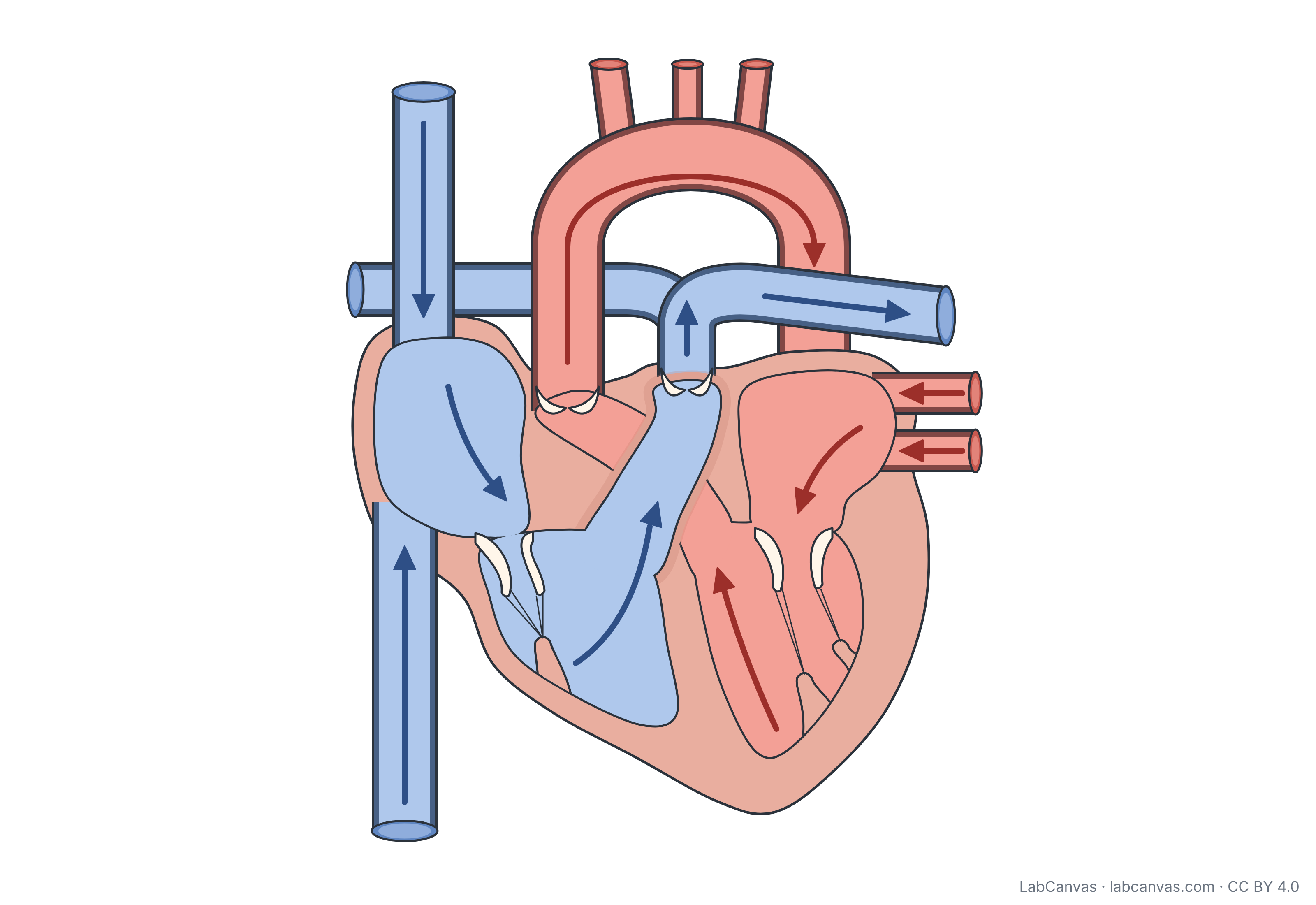
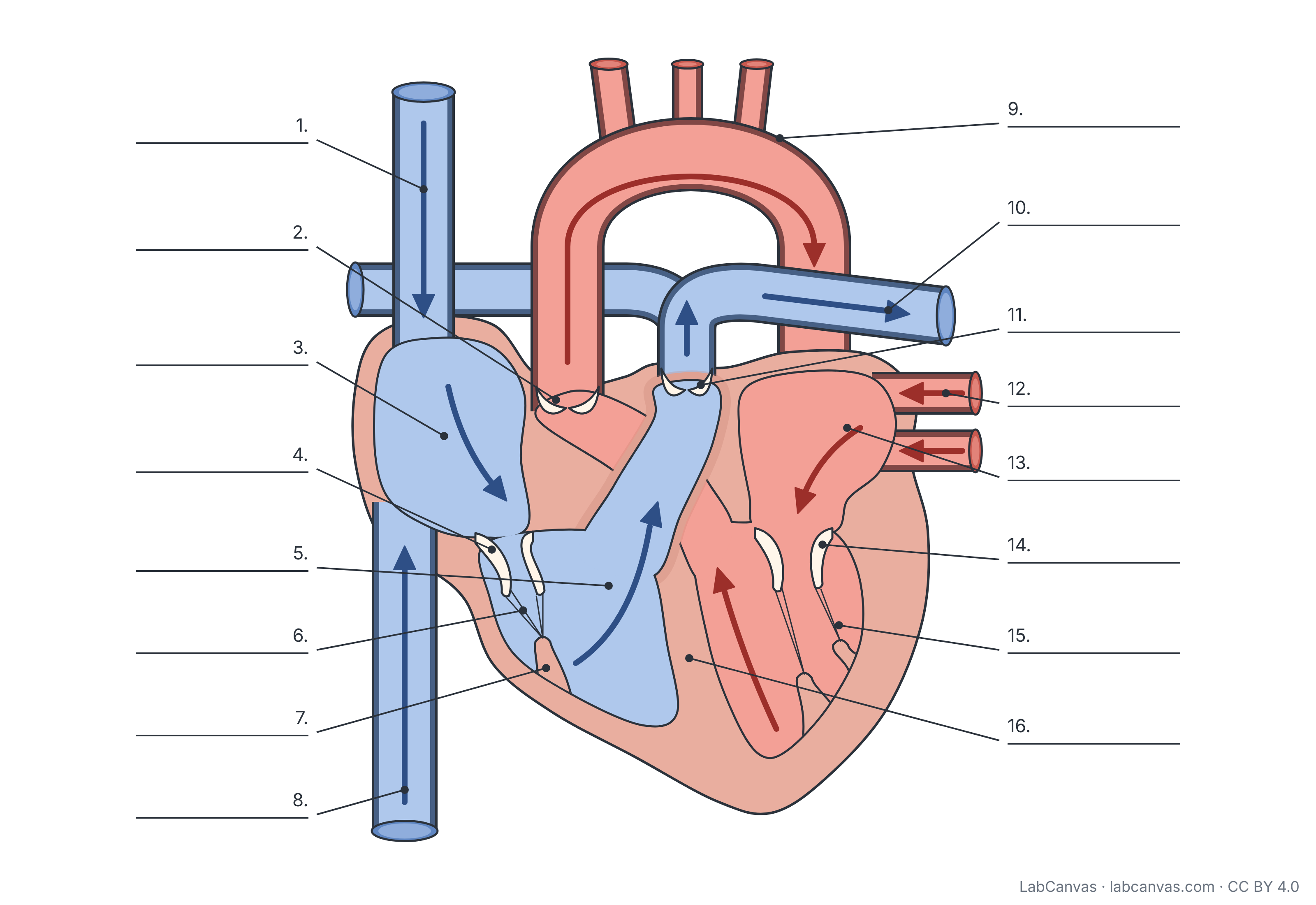
The human heart has four chambers: two upper atria that receive blood and two lower ventricles that pump it out. The right side sends deoxygenated blood to the lungs through the pulmonary artery, and the left side sends oxygenated blood to the body through the aorta. Four valves (tricuspid, pulmonary, mitral and aortic) keep blood flowing one way. This free diagram labels 16 parts and comes unlabelled and as a numbered worksheet.
Last updated
Download the heart diagram
Free to use in homework, teaching, slides, posters and publications under CC BY 4.0. Please credit: Heart diagram by LabCanvas (labcanvas.com), CC BY 4.0
Parts of the heart and what they do
| # | Part | What it is or does |
|---|---|---|
| 1 | Superior vena cava | Large vein that returns deoxygenated blood from the head, neck, arms and chest to the right atrium. |
| 2 | Aortic valve | Semilunar valve between the left ventricle and the aorta; it opens when the ventricle contracts and closes to stop blood flowing back. |
| 3 | Right atrium | Upper right chamber; receives deoxygenated blood from the body through the venae cavae and passes it to the right ventricle. |
| 4 | Tricuspid valve | Right atrioventricular valve with three flaps (cusps); lets blood pass from the right atrium to the right ventricle but not back. |
| 5 | Right ventricle | Lower right chamber; pumps deoxygenated blood through the pulmonary valve into the pulmonary trunk towards the lungs. |
| 6 | Chordae tendineae | Strong tendon-like cords that tie the flaps of the atrioventricular valves to the papillary muscles. |
| 7 | Papillary muscle | Cone of ventricle muscle that tightens the chordae tendineae as the ventricle contracts, so the valve flaps cannot turn back into the atrium. |
| 8 | Inferior vena cava | Large vein that returns deoxygenated blood from the lower body (abdomen, pelvis and legs) to the right atrium. |
| 9 | Aorta | The largest artery; carries oxygenated blood from the left ventricle and, through its branches, to the whole body. |
| 10 | Pulmonary artery | The pulmonary trunk and its left and right branches; carry deoxygenated blood from the right ventricle to the lungs. |
| 11 | Pulmonary valve | Semilunar valve between the right ventricle and the pulmonary trunk; stops blood flowing back into the ventricle. |
| 12 | Pulmonary veins | Veins that bring oxygenated blood from the lungs into the left atrium. |
| 13 | Left atrium | Upper left chamber; receives oxygenated blood from the pulmonary veins and passes it to the left ventricle. |
| 14 | Mitral (bicuspid) valve | Left atrioventricular valve with two flaps; lets blood pass from the left atrium to the left ventricle but not back. |
| 15 | Left ventricle | Lower left chamber with the thickest muscular wall; pumps oxygenated blood through the aortic valve into the aorta. |
| 16 | Interventricular septum | Thick wall of heart muscle that separates the right and left ventricles so the two blood streams do not mix. |
How to label a heart diagram
- Get your bearingsThe diagram is drawn as if you face the person, so the heart's right side is on your left. Blue marks deoxygenated blood and red marks oxygenated blood.
- Label the four chambersThe two thin-walled upper chambers are the right and left atria; the two lower chambers are the right and left ventricles. The left ventricle has the thickest wall.
- Add the septumLabel the thick muscle wall between the two ventricles as the interventricular septum.
- Follow blood into the heartThe superior vena cava enters the right atrium from above and the inferior vena cava from below. The pulmonary veins enter the left atrium from the lungs.
- Follow blood out of the heartThe pulmonary artery leaves the right ventricle and branches to both lungs. The aorta leaves the left ventricle and arches over the top of the heart.
- Label the valvesTricuspid: right atrium to right ventricle. Pulmonary: right ventricle to pulmonary artery. Mitral (bicuspid): left atrium to left ventricle. Aortic: left ventricle to aorta. Finish with the chordae tendineae and papillary muscles under the tricuspid valve, then check against the key.
Right side vs left side of the heart
| Feature | Right side | Left side |
|---|---|---|
| Blood it carries | Deoxygenated blood from the body | Oxygenated blood from the lungs |
| Atrium receives blood from | Superior and inferior vena cava (and the coronary sinus) | Pulmonary veins |
| Atrioventricular valve | Tricuspid valve (three flaps) | Mitral or bicuspid valve (two flaps) |
| Semilunar valve | Pulmonary valve | Aortic valve |
| Ventricle pumps blood into | Pulmonary trunk, to the lungs | Aorta, to the rest of the body |
| Circuit it serves | Pulmonary circuit | Systemic circuit |
| Ventricle wall | Thinner | Much thicker, because it pumps blood around the whole body |
The path of blood through the heart
Blood follows one loop through the heart and lungs and a second loop through the body. Deoxygenated blood from the body enters the right atrium through the superior and inferior venae cavae, passes the tricuspid valve into the right ventricle, and is pumped through the pulmonary valve into the pulmonary trunk and pulmonary arteries to the lungs, where it picks up oxygen and gives up carbon dioxide. This is the pulmonary circuit.
Oxygenated blood returns through the pulmonary veins to the left atrium, passes the mitral (bicuspid) valve into the left ventricle, and is pumped through the aortic valve into the aorta, which carries it to the rest of the body. This is the systemic circuit. Both atria contract together, then both ventricles contract together.
How the heart valves work
The valves open and close because of differences in pressure, which keeps blood moving in one direction. The two atrioventricular valves (tricuspid and mitral) sit between each atrium and ventricle. When the ventricles contract, the rising pressure pushes their flaps shut, and the papillary muscles pull on the chordae tendineae so the flaps cannot be forced back into the atria.
The two semilunar valves (pulmonary and aortic) sit where the arteries leave the ventricles. Their pocket-shaped cusps open as blood is pumped out and fill and close when the ventricles relax. The closing of the valves makes the familiar heart sounds.
Why is one side blue and the other red?
Diagrams colour deoxygenated blood blue and oxygenated blood red only to make the two circuits easy to follow. Real blood is always red: oxygen-rich blood is bright red and oxygen-poor blood is a darker red. Note that the pulmonary arteries carry deoxygenated blood and the pulmonary veins carry oxygenated blood, the reverse of the other arteries and veins, because arteries are defined as vessels that carry blood away from the heart and veins as vessels that carry it back.
Quick quiz
1. Which chamber receives blood from the superior and inferior vena cava?
- Left atrium
- Right atrium
- Right ventricle
- Left ventricle
Answer: b) Right atrium. The venae cavae return deoxygenated blood from the body to the right atrium.
2. Which valve lies between the left atrium and the left ventricle?
- Tricuspid valve
- Aortic valve
- Mitral (bicuspid) valve
- Pulmonary valve
Answer: c) Mitral (bicuspid) valve. The mitral (bicuspid) valve is the left atrioventricular valve.
3. Which vessel carries oxygenated blood from the lungs to the heart?
- Pulmonary artery
- Aorta
- Superior vena cava
- Pulmonary veins
Answer: d) Pulmonary veins. The pulmonary veins return oxygenated blood from the lungs to the left atrium.
4. Which chamber has the thickest muscular wall?
- Left ventricle
- Right ventricle
- Left atrium
- Right atrium
Answer: a) Left ventricle. The left ventricle pumps blood around the whole body, so it has the thickest wall.
5. What separates the right and left ventricles?
- Chordae tendineae
- Interventricular septum
- Pulmonary valve
- Papillary muscle
Answer: b) Interventricular septum. The interventricular septum is the muscular wall between the two ventricles.
Frequently asked questions
What are the 4 chambers of the heart?
The right atrium, right ventricle, left atrium and left ventricle. The atria are the upper chambers that receive blood; the ventricles are the lower chambers that pump blood out of the heart.
What are the 4 valves of the heart?
The tricuspid valve (right atrium to right ventricle), the pulmonary valve (right ventricle to pulmonary trunk), the mitral or bicuspid valve (left atrium to left ventricle) and the aortic valve (left ventricle to aorta).
What is the order of blood flow through the heart?
Venae cavae, right atrium, tricuspid valve, right ventricle, pulmonary valve, pulmonary arteries, lungs, pulmonary veins, left atrium, mitral valve, left ventricle, aortic valve, aorta, body.
Why is the left ventricle wall thicker than the right?
The left ventricle pumps blood around the whole body through the systemic circuit, which takes much more force than pumping blood the short distance to the lungs, so its muscular wall is much thicker.
Do pulmonary arteries carry oxygenated blood?
No. The pulmonary arteries carry deoxygenated blood from the right ventricle to the lungs, and the pulmonary veins carry oxygenated blood back to the left atrium. Arteries are named for carrying blood away from the heart, not for their oxygen content.
Why is the heart diagram drawn back to front?
Heart diagrams show the heart as you would see it facing a person, so the heart's right side appears on your left. That is why the right atrium and right ventricle are drawn on the left of the picture.
What do the chordae tendineae do?
They are tendon-like cords that link the flaps of the tricuspid and mitral valves to the papillary muscles. When the ventricles contract, they hold the flaps in place so blood cannot leak back into the atria.
Sources
- OpenStax Anatomy and Physiology 2e, 19.1 Heart Anatomy (CC BY 4.0)
- OpenStax Anatomy and Physiology 2e, 19.3 Cardiac Cycle (CC BY 4.0)
Facts on this page follow these introductory textbooks (OpenStax, CC BY 4.0). The diagram is a simplified illustration; organelles are not drawn to scale.
More free diagrams
Make your own science diagrams
LabCanvas is a browser workspace for scientific figures, slides and posters, with adjustable cells, organelles and molecules.
Edit this diagram