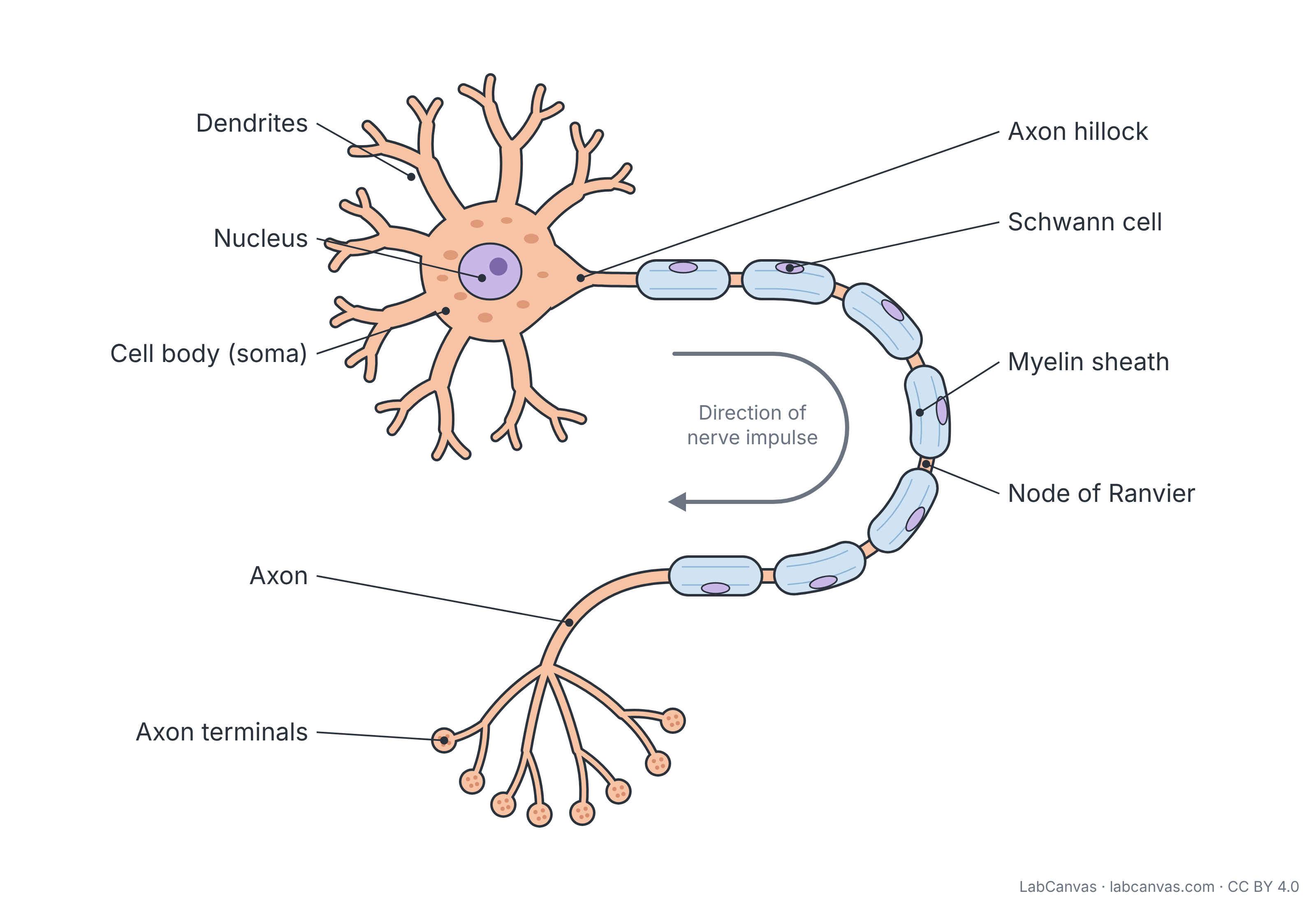
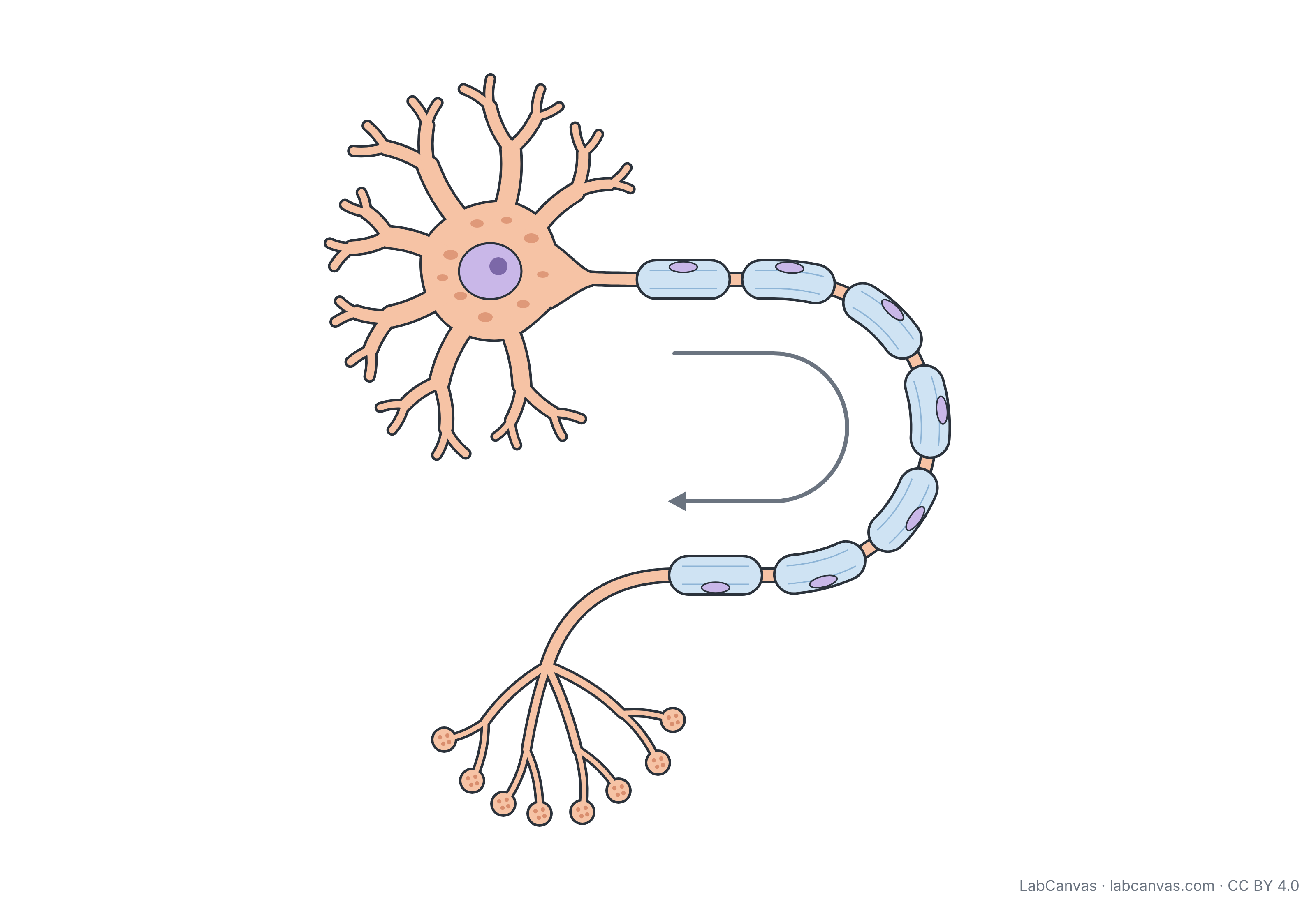
Neuron diagram with labels
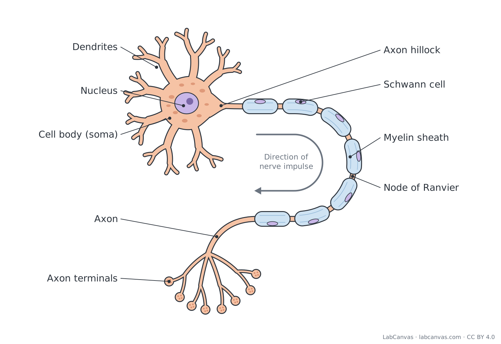
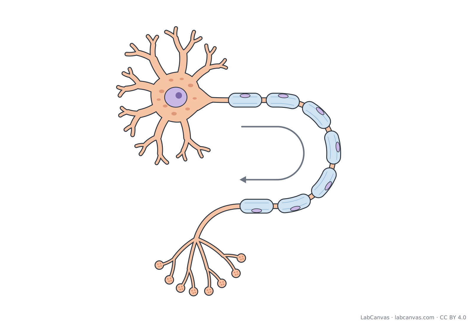
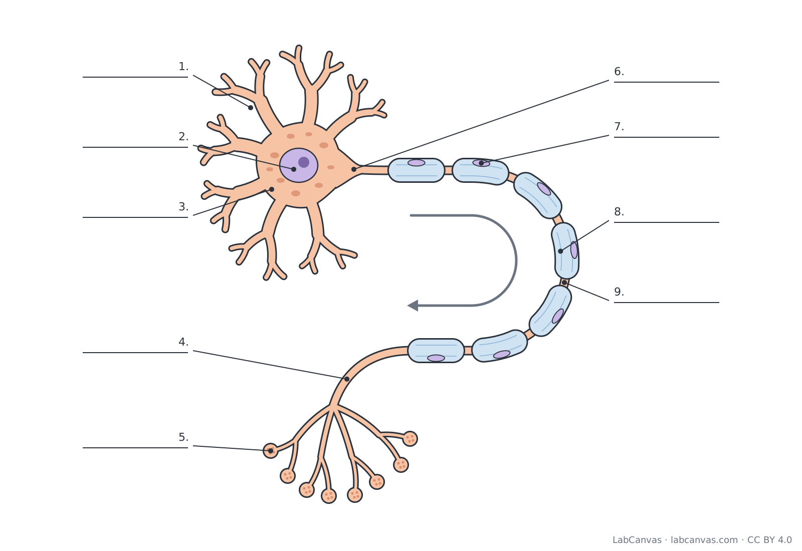
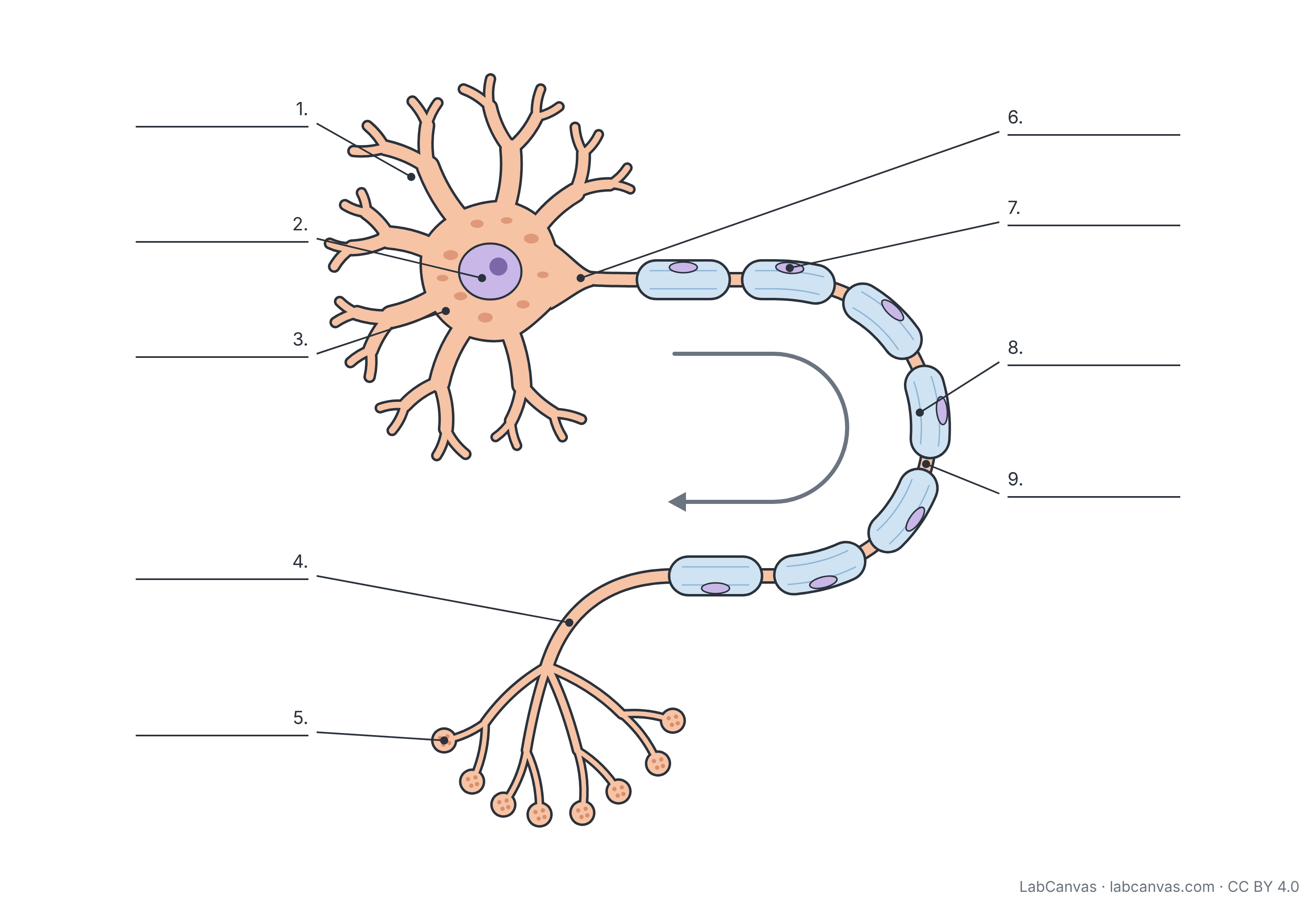
A neuron is a nerve cell that carries electrical and chemical signals. Its dendrites receive signals and pass them to the cell body (soma), which holds the nucleus; the signal then travels from the axon hillock along a single axon, insulated by a myelin sheath with gaps called nodes of Ranvier, to the axon terminals, which pass it on to the next cell. This free diagram of a multipolar motor neuron labels 9 parts and comes unlabelled and as a numbered worksheet with an answer key.
Last updated
Download the neuron diagram
Free to use in homework, teaching, slides, posters and publications under CC BY 4.0. Please credit: Neuron diagram by LabCanvas (labcanvas.com), CC BY 4.0
Parts of the neuron and what they do
| # | Part | What it is or does |
|---|---|---|
| 1 | Dendrites | Short, branched extensions of the cell body that receive signals from other neurons and carry them towards the cell body. |
| 2 | Nucleus | Holds the neuron's DNA and controls the cell; it sits in the cell body. |
| 3 | Cell body (soma) | The main part of the neuron, containing the nucleus and most organelles; it combines the incoming signals and keeps the cell alive. |
| 4 | Axon | A single long extension that carries the nerve impulse (action potential) away from the cell body towards other cells. |
| 5 | Axon terminals | Branched endings of the axon (synaptic terminals); they release neurotransmitter at synapses to pass the signal to another neuron, muscle or gland. |
| 6 | Axon hillock | Cone-shaped region where the axon leaves the cell body; signals are added up here, and if they are strong enough an action potential starts. |
| 7 | Schwann cell | Glial cell of the peripheral nervous system that wraps around a section of the axon to form the myelin sheath; its nucleus lies on the outside. |
| 8 | Myelin sheath | Fatty insulating layers of glial cell membrane wrapped around the axon; it makes the nerve impulse travel much faster. |
| 9 | Node of Ranvier | Short gap in the myelin sheath between two glial cells where the axon is exposed; the impulse jumps from node to node (saltatory conduction). |
How to label a neuron diagram
- Find the cell bodyStart with the large rounded part with a purple nucleus inside. That is the cell body (soma), and the nucleus is in its centre.
- Label the dendritesThe many short, tree-like branches growing out of the cell body are dendrites. They bring signals in.
- Follow the axonFind the one long fibre leaving the cell body. Where it narrows into a cone is the axon hillock, and the fibre itself is the axon.
- Label the insulationThe blue sausage-shaped segments along the axon form the myelin sheath. Each one is made by a Schwann cell, whose nucleus shows on the outside. The bare gaps between segments are the nodes of Ranvier.
- Finish at the end of the axonThe small branches with round knobs at the end of the axon are the axon terminals. Follow the arrow to check the direction of the impulse, then check your answers against the key.
Dendrites vs axon
| Feature | Dendrites | Axon |
|---|---|---|
| Number per neuron | Usually many | Usually one (it may branch at its end) |
| Direction of signal | Towards the cell body | Away from the cell body |
| Shape | Short and highly branched, tapering | Long and thin, of even width |
| Myelin sheath | No | Often (myelinated axons) |
| Ends in | Fine branches near the cell body | Axon terminals that form synapses |
How a signal travels through a neuron
Signals from other neurons arrive at the dendrites and the cell body. They are added together at the axon hillock; if the combined signal is strong enough, an action potential (nerve impulse) starts and travels along the axon. In a myelinated axon the impulse jumps from one node of Ranvier to the next, which is much faster than moving along a bare axon. At the axon terminals the impulse causes chemical messengers called neurotransmitters to be released into the synapse, where they act on the next neuron, a muscle fibre or a gland.
Myelin: Schwann cells and oligodendrocytes
Myelin is made by glial cells, the support cells of the nervous system. In the peripheral nervous system (the nerves outside the brain and spinal cord) each Schwann cell wraps around one section of one axon. In the central nervous system oligodendrocytes do the same job, and one oligodendrocyte can myelinate sections of several axons. Damage to myelin slows or blocks nerve signals, which is what happens in multiple sclerosis.
Types of neurons
Neurons are grouped by shape. Multipolar neurons, such as the motor neuron shown here, have one axon and many dendrites. Bipolar neurons have one axon and one dendrite, as in the retina. Unipolar and pseudounipolar neurons have a single process from the cell body; many sensory neurons are pseudounipolar. By job, sensory neurons carry information to the central nervous system, motor neurons carry commands from it to muscles and glands, and interneurons connect neurons within it.
Quick quiz
1. Which part of a neuron receives signals from other neurons?
- Axon terminals
- Dendrites
- Myelin sheath
- Node of Ranvier
Answer: b) Dendrites. Dendrites receive incoming signals and carry them towards the cell body.
2. Which cells make the myelin sheath in the peripheral nervous system?
- Oligodendrocytes
- Astrocytes
- Schwann cells
- Red blood cells
Answer: c) Schwann cells. Schwann cells wrap around axons in the peripheral nervous system; oligodendrocytes do this in the brain and spinal cord.
3. What are the gaps in the myelin sheath called?
- Synapses
- Nodes of Ranvier
- Axon hillocks
- Dendrites
Answer: b) Nodes of Ranvier. Nodes of Ranvier are the exposed gaps between myelinated sections.
4. Where does an action potential usually start?
- Axon hillock
- Axon terminal
- Nucleus
- Dendrite tip
Answer: a) Axon hillock. Signals are added up at the axon hillock, where an action potential starts if the threshold is reached.
5. In which direction does the impulse travel along the axon?
- Towards the cell body
- Away from the cell body
- Both ways equally
- Only between dendrites
Answer: b) Away from the cell body. The axon carries the impulse away from the cell body towards the axon terminals.
Frequently asked questions
What are the parts of a neuron?
A typical neuron has dendrites, a cell body (soma) containing the nucleus, an axon hillock, an axon (often covered by a myelin sheath with gaps called nodes of Ranvier) and axon terminals that form synapses with other cells.
What is the function of the myelin sheath?
The myelin sheath insulates the axon so that the nerve impulse jumps from one node of Ranvier to the next. This makes signals travel much faster than along an unmyelinated axon.
What is the difference between dendrites and axons?
Dendrites are short, branched and usually many; they carry signals towards the cell body. The axon is usually single and long; it carries signals away from the cell body to other cells.
What are nodes of Ranvier?
Nodes of Ranvier are small gaps in the myelin sheath between neighbouring glial cells. The axon membrane is exposed there, and the action potential jumps from node to node (saltatory conduction).
What is the axon hillock?
The axon hillock is the cone-shaped region where the axon joins the cell body. Incoming signals are added up there, and if they reach the threshold an action potential begins.
What do axon terminals do?
Axon terminals release neurotransmitters into the synapse. The neurotransmitters bind to receptors on the next neuron, muscle cell or gland cell and pass the signal on.
Is a motor neuron multipolar?
Yes. A motor neuron is a multipolar neuron: it has many dendrites and a single long axon that carries commands from the brain or spinal cord to a muscle.
Sources
- OpenStax Biology 2e, 35.1 Neurons and Glial Cells (CC BY 4.0)
- OpenStax Anatomy and Physiology 2e, 12.2 Nervous Tissue (CC BY 4.0)
Facts on this page follow these introductory textbooks (OpenStax, CC BY 4.0). The diagram is a simplified illustration; organelles are not drawn to scale.
More free diagrams
Make your own science diagrams
LabCanvas is a browser workspace for scientific figures, slides and posters, with adjustable cells, organelles and molecules.
Edit this diagram