Digestive system diagram with labels
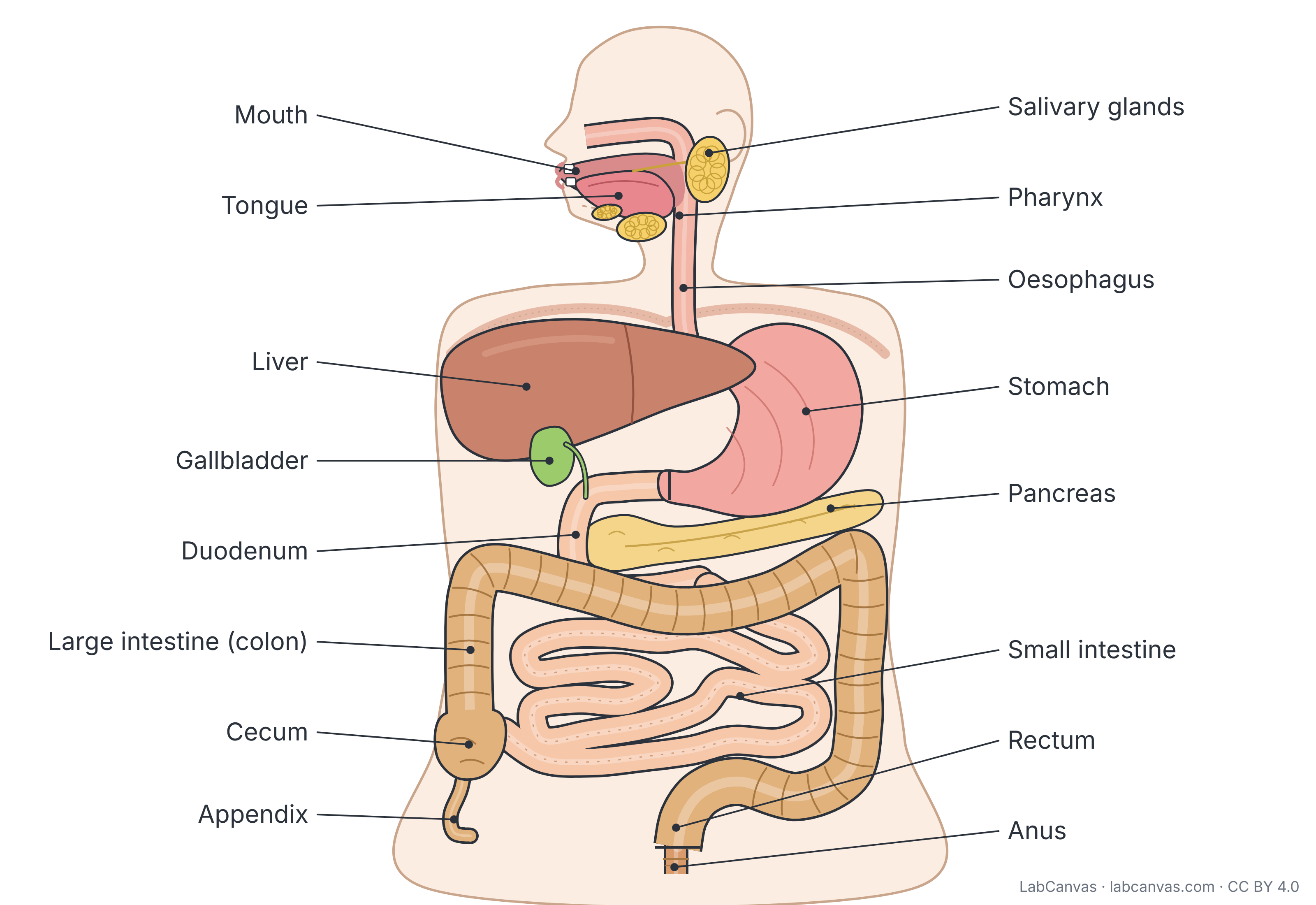
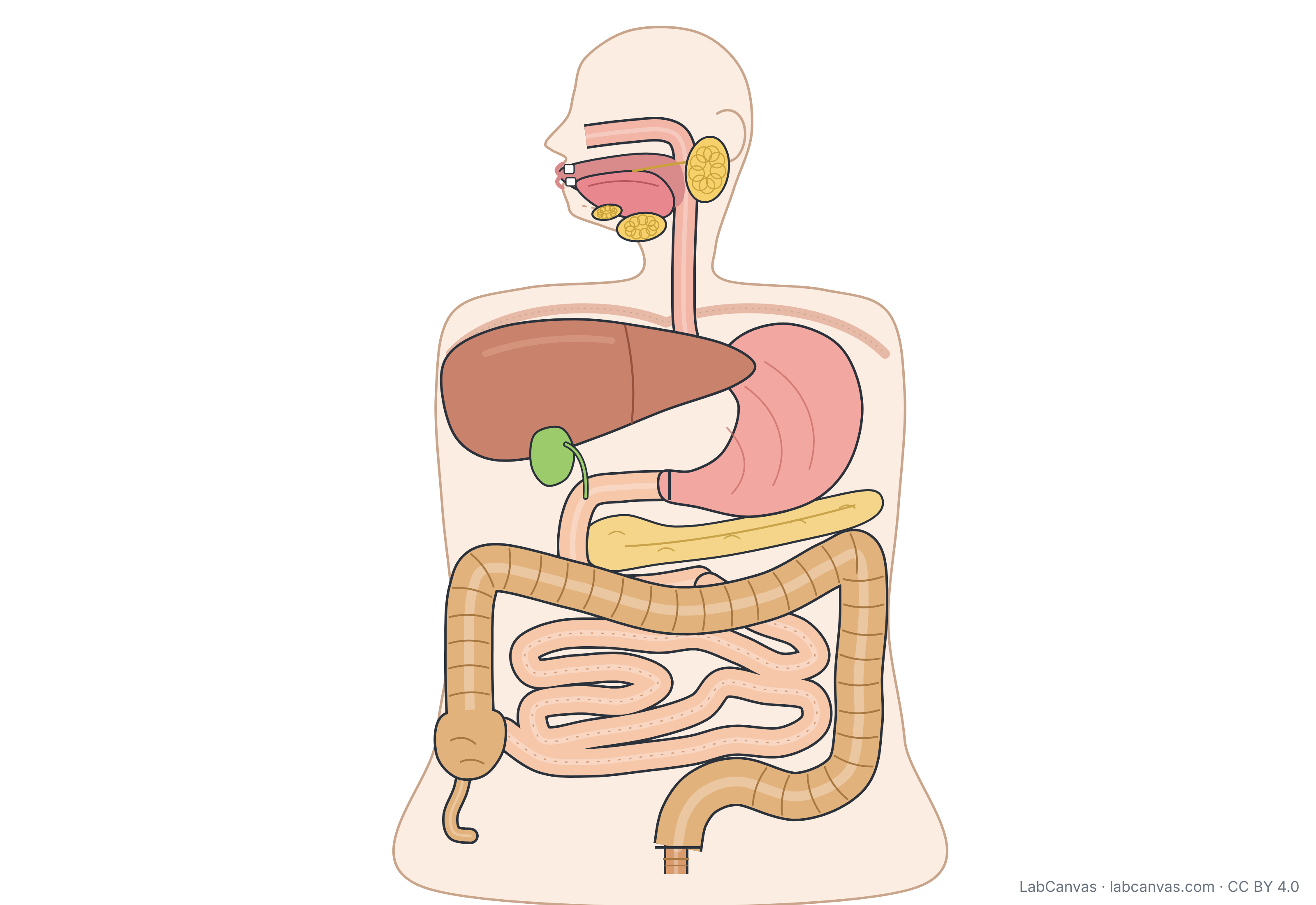
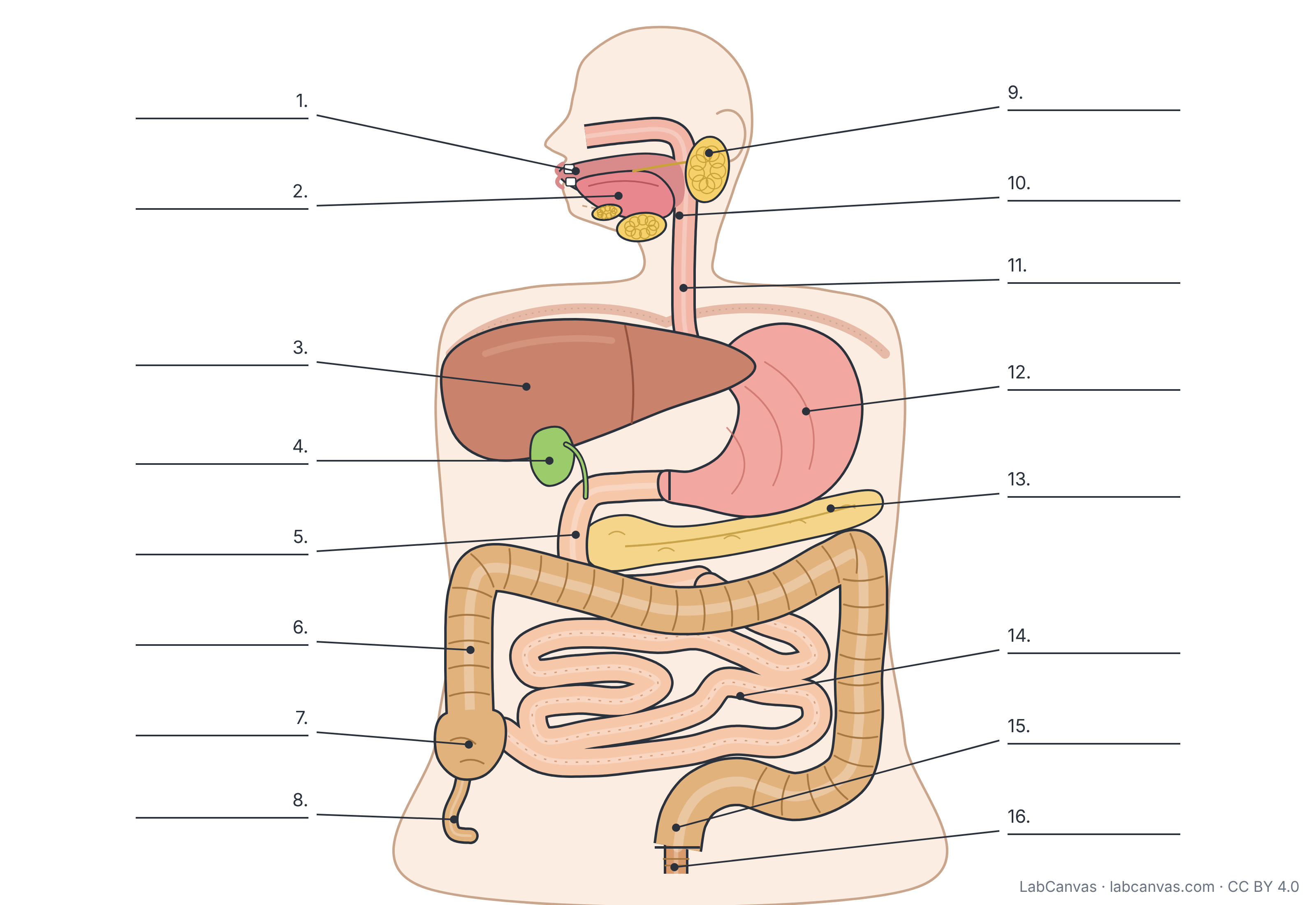
The digestive system is the alimentary canal, one continuous tube from the mouth through the pharynx, oesophagus, stomach, small intestine and large intestine to the anus, plus accessory organs that help digestion: the teeth, tongue, salivary glands, liver, gallbladder and pancreas. Food is broken down mechanically and chemically, nutrients are absorbed mainly in the small intestine, and waste leaves as feces. This free diagram labels 16 parts and comes unlabelled and as a numbered worksheet.
Last updated
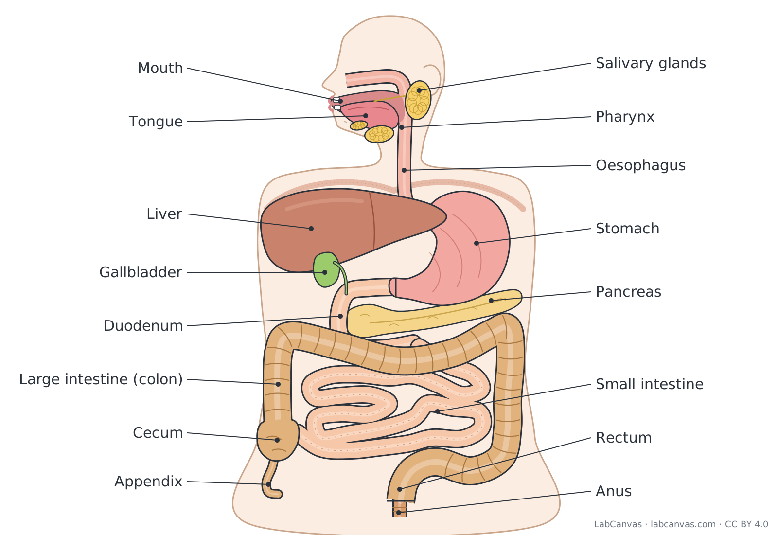
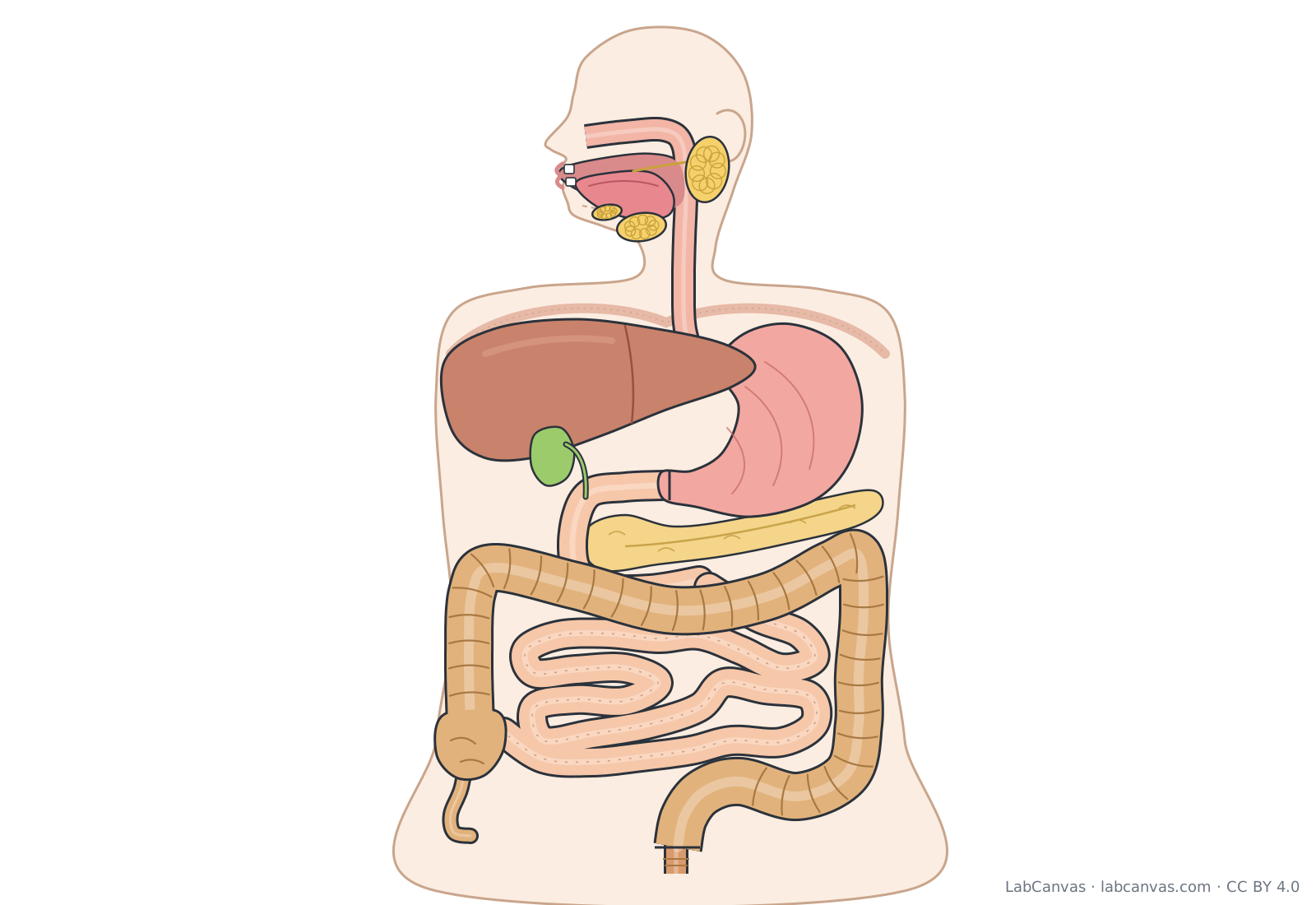
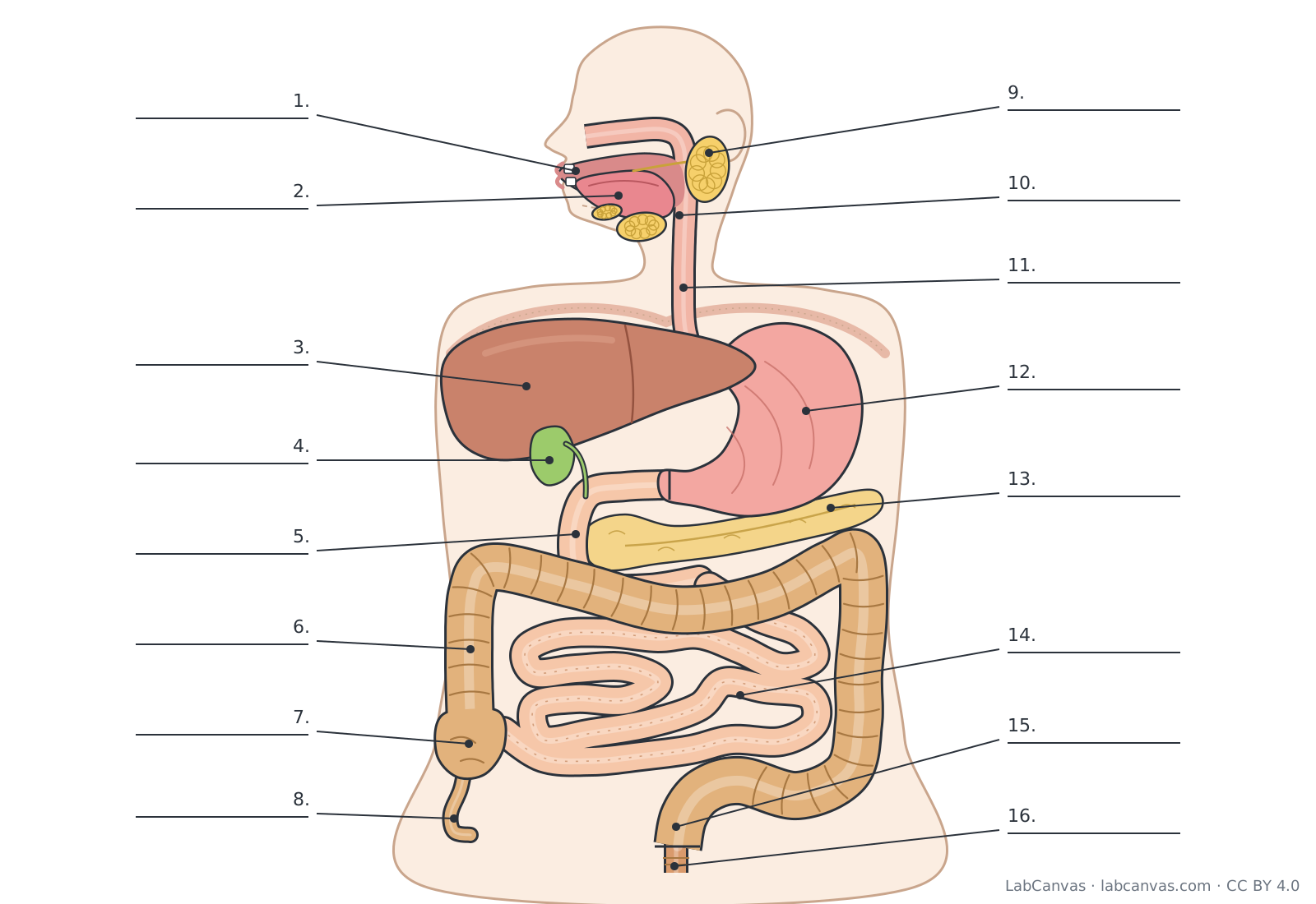
Download the digestive system diagram
Free to use in homework, teaching, slides, posters and publications under CC BY 4.0. Please credit: Digestive system diagram by LabCanvas (labcanvas.com), CC BY 4.0
Parts of the digestive system and what they do
| # | Part | What it is or does |
|---|---|---|
| 1 | Mouth | Where digestion begins: the teeth chew food (mechanical digestion) and saliva starts chemical digestion of starch. |
| 2 | Tongue | Muscular organ that mixes food with saliva, shapes it into a soft ball (bolus) and pushes it back for swallowing. |
| 3 | Liver | Large organ in the upper right abdomen; makes bile, which helps break fats into small droplets, and processes absorbed nutrients. |
| 4 | Gallbladder | Small sac under the liver that stores and concentrates bile and releases it into the duodenum. |
| 5 | Duodenum | First, C-shaped part of the small intestine; receives chyme from the stomach, bile and pancreatic juice. |
| 6 | Large intestine (colon) | Wider final part of the tract (cecum, colon, rectum, anal canal); absorbs water, houses gut bacteria and forms feces. |
| 7 | Cecum | Pouch at the start of the large intestine, where the ileum of the small intestine empties. |
| 8 | Appendix | Narrow, worm-like tube attached to the cecum; contains lymphoid tissue. |
| 9 | Salivary glands | Three pairs of glands (parotid, submandibular, sublingual) that release saliva into the mouth to moisten food and begin digesting starch. |
| 10 | Pharynx | Throat; a passage shared by food and air that carries swallowed food from the mouth to the oesophagus. |
| 11 | Oesophagus | Muscular tube (esophagus) that moves food from the pharynx to the stomach by waves of muscle contraction called peristalsis. |
| 12 | Stomach | J-shaped muscular sac that stores food, churns it with acid and enzymes such as pepsin into chyme, and starts protein digestion. |
| 13 | Pancreas | Gland behind the stomach; sends digestive enzymes and bicarbonate to the duodenum and makes hormones such as insulin. |
| 14 | Small intestine | Long, coiled tube (duodenum, jejunum, ileum) where most digestion and almost all nutrient absorption take place. |
| 15 | Rectum | Final straight part of the large intestine that stores feces until they are eliminated. |
| 16 | Anus | Opening at the end of the anal canal, controlled by sphincter muscles, through which feces leave the body. |
How to label a digestive system diagram
- Start at the mouthIn the head, label the mouth, the tongue on its floor, and the salivary glands: one in front of the ear and two under the jaw and tongue.
- Follow the tube downThe passage behind the mouth is the pharynx; it continues down the neck and chest as the oesophagus, which ends at the stomach under the diaphragm.
- Label the upper abdomenThe large organ on the left of the picture (the body's right side) is the liver, with the small green gallbladder under it. The J-shaped sac on the right of the picture is the stomach, and the long gland below it is the pancreas.
- Trace the small intestineThe C-shaped tube leaving the stomach is the duodenum; the tightly coiled tube in the middle of the abdomen is the rest of the small intestine.
- Trace the large intestineStart at the cecum, the pouch at the bottom of the picture's left side, with the appendix hanging from it. Follow the colon up, across and down around the small intestine.
- Finish at the end of the tractThe colon leads into the rectum and the short anal canal, which opens at the anus. Check your labels against the answer key.
Small intestine vs large intestine
| Feature | Small intestine | Large intestine |
|---|---|---|
| Parts | Duodenum, jejunum, ileum | Cecum (with appendix), colon, rectum, anal canal |
| Width and length | Narrower, but much longer | Wider, but shorter |
| Main job | Finishes digestion and absorbs almost all nutrients | Absorbs water and forms and stores feces |
| Inner surface | Folds, villi and microvilli give a huge absorbing surface | No villi; pouches called haustra in the colon wall |
| Receives | Chyme from the stomach, bile and pancreatic juice | What is left from the ileum |
The path food takes
The alimentary canal (gastrointestinal or GI tract) is one continuous tube. Food is chewed in the mouth and mixed with saliva, swallowed through the pharynx, and carried down the oesophagus by peristalsis to the stomach. The stomach churns it into a liquid called chyme and releases it a little at a time into the duodenum, the first part of the small intestine, which continues as the jejunum and ileum.
What is not absorbed passes into the cecum of the large intestine and through the ascending, transverse, descending and sigmoid colon to the rectum, and leaves through the anal canal and anus.
Accessory digestive organs
Food never passes through the accessory organs, but they are essential to digestion. The teeth and tongue break up and move food, and the salivary glands add saliva, which contains an enzyme (salivary amylase) that starts digesting starch. The liver makes bile, which emulsifies fats; the gallbladder stores and concentrates it. The pancreas produces pancreatic juice, full of digestive enzymes and bicarbonate that neutralises acid chyme. Bile and pancreatic juice both empty into the duodenum.
Mechanical and chemical digestion
Mechanical digestion breaks food into smaller pieces without changing its chemistry: chewing in the mouth, churning in the stomach and segmentation in the small intestine. Chemical digestion uses enzymes to split large food molecules into small ones the body can absorb: carbohydrates into simple sugars, proteins into amino acids, and fats into fatty acids and monoglycerides. Chemical digestion starts in the mouth and is completed in the small intestine, where the nutrients are absorbed into the blood and lymph.
Quick quiz
1. Which organ makes bile?
- Gallbladder
- Pancreas
- Liver
- Stomach
Answer: c) Liver. The liver makes bile; the gallbladder only stores and concentrates it.
2. What is the first part of the small intestine called?
- Ileum
- Duodenum
- Jejunum
- Cecum
Answer: b) Duodenum. The duodenum is the first, C-shaped part of the small intestine.
3. Which tube carries food from the pharynx to the stomach?
- Trachea
- Oesophagus
- Colon
- Duodenum
Answer: b) Oesophagus. The oesophagus moves food to the stomach by peristalsis.
4. Where are most nutrients absorbed?
- Stomach
- Large intestine
- Small intestine
- Oesophagus
Answer: c) Small intestine. Almost all nutrient absorption happens in the small intestine.
5. Which of these is an accessory organ, not part of the alimentary canal?
- Pancreas
- Stomach
- Rectum
- Pharynx
Answer: a) Pancreas. The pancreas releases juice into the duodenum, but food never passes through it.
Frequently asked questions
What are the organs of the digestive system?
The alimentary canal: mouth, pharynx, oesophagus, stomach, small intestine (duodenum, jejunum, ileum) and large intestine (cecum, colon, rectum, anal canal) ending at the anus. The accessory organs are the teeth, tongue, salivary glands, liver, gallbladder and pancreas.
What is the order of the digestive system?
Mouth, pharynx, oesophagus, stomach, small intestine (duodenum, jejunum, ileum), large intestine (cecum, ascending, transverse, descending and sigmoid colon), rectum, anal canal and anus.
Where does most digestion happen?
In the small intestine. Enzymes from the pancreas and the intestinal wall, helped by bile from the liver, finish digesting carbohydrates, proteins and fats there, and almost all nutrients are absorbed through its wall.
What does the large intestine do?
It absorbs water and some salts from the remaining material, hosts large numbers of gut bacteria that break down some leftover substances, and forms and stores feces until they are eliminated.
Does food pass through the liver, pancreas or gallbladder?
No. These are accessory organs. They release bile and pancreatic juice into the duodenum through ducts, but food itself only travels through the alimentary canal.
What is the difference between the oesophagus and the pharynx?
The pharynx (throat) is the passage behind the mouth and nose shared by food and air. The oesophagus (esophagus) is the muscular tube below it that carries only food, from the pharynx to the stomach.
What does the appendix do?
The appendix is a narrow tube attached to the cecum. It contains lymphoid tissue, which is part of the immune system; it does not play a direct part in digesting food.
Sources
- OpenStax Anatomy and Physiology 2e, 23.1 Overview of the Digestive System (CC BY 4.0)
- OpenStax Anatomy and Physiology 2e, 23.3 The Mouth, Pharynx, and Esophagus (CC BY 4.0)
- OpenStax Anatomy and Physiology 2e, 23.4 The Stomach (CC BY 4.0)
- OpenStax Anatomy and Physiology 2e, The Small and Large Intestines (CC BY 4.0)
- OpenStax Anatomy and Physiology 2e, Accessory Organs in Digestion: The Liver, Pancreas, and Gallbladder (CC BY 4.0)
Facts on this page follow these introductory textbooks (OpenStax, CC BY 4.0). The diagram is a simplified illustration; organelles are not drawn to scale.
More free diagrams
Make your own science diagrams
LabCanvas is a browser workspace for scientific figures, slides and posters, with adjustable cells, organelles and molecules.
Edit this diagram