The LabCanvas Protein viewer: PDB structures styled for figures
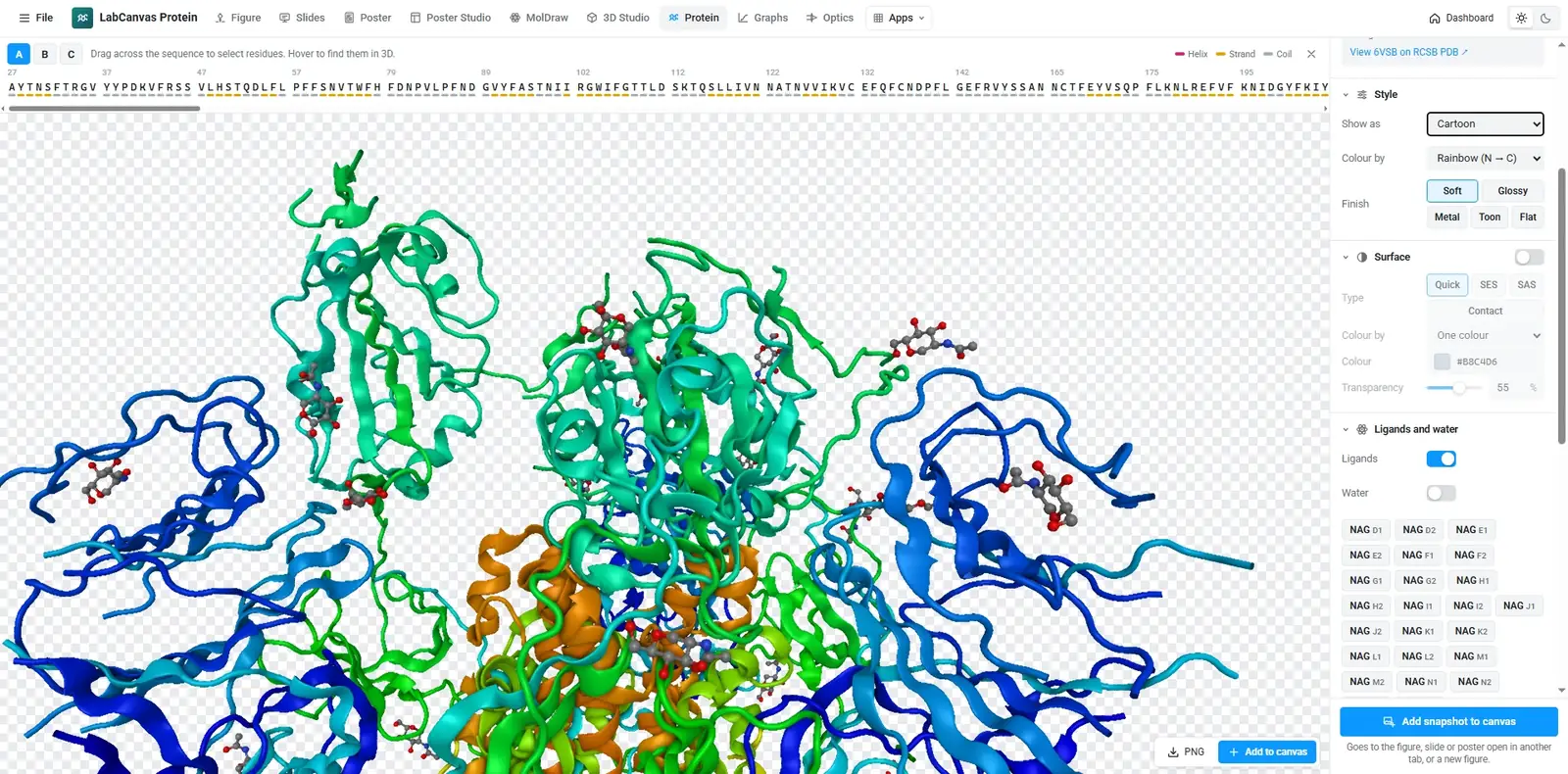
How to show a protein structure in the LabCanvas Protein viewer: load any PDB ID or your own PDB and mmCIF file, choose cartoon, surface or ball-and-stick, colour by chain or secondary structure, select residues on the sequence and snapshot a transparent PNG for your figure.
Contents
The LabCanvas Protein viewer is a focused viewer for protein structures. Load any entry of the RCSB Protein Data Bank by its ID, or your own PDB or mmCIF file, choose a style and a colouring, select residues on the sequence to give them their own look, and snapshot a transparent PNG straight into your figure.
What it is for
- Structure panels in papers: a cartoon coloured by chain with the active site in ball-and-stick.
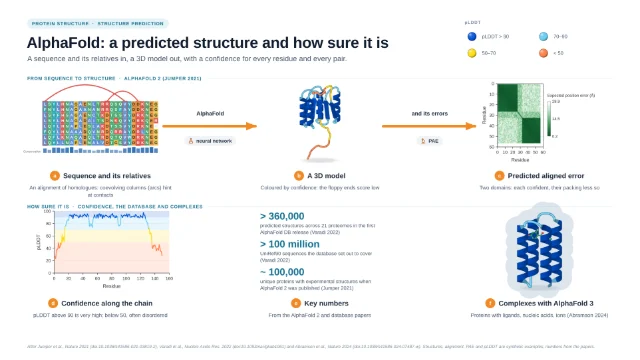
- Predicted models coloured by confidence (AlphaFold writes pLDDT into the B-factor column), next to the PAE plot made in Graphs & statistics.
- Teaching figures of secondary structure, domains and binding sites.
Styles and colourings
| Option | Choices |
|---|---|
| Representation | Cartoon, tube, trace, ball-and-stick, sticks, space-filling, lines, quick, solvent-excluded and solvent-accessible surfaces |
| Colouring | Secondary structure, chain, rainbow (N → C), element, residue type, hydrophobicity, B-factor or one colour |
| Materials | Soft, glossy, metal, toon or flat |
| Selection | Residues picked on the sequence bar get their own style and colour |
| Output | Transparent PNG snapshot into a figure, slide or poster |
How to show a protein and highlight residues
- Open the Protein viewer and enter a PDB ID such as 4HHB.
- Pick a style and colouring, for example cartoon by chain.
- Drag across residues on the sequence bar to select them.
- Give the selection its own style (ball-and-stick) and colour.
- Turn the view, then take a snapshot and add it to your figure.
Protein figures made in LabCanvas
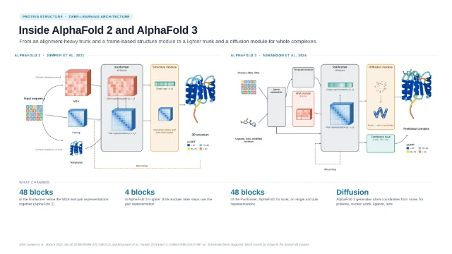
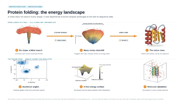
The template library includes graphical abstracts of protein structure prediction, built from protein objects, confidence plots and diagrams you can edit.
Frequently asked questions
Which structures can I open?
Which styles and colourings are there?
How do I highlight a binding site?
How do I put the protein in a figure?
What powers the viewer?
Try it in LabCanvas
Every app is free to start in your browser. Create an account and begin.
Open Protein viewerKeep reading
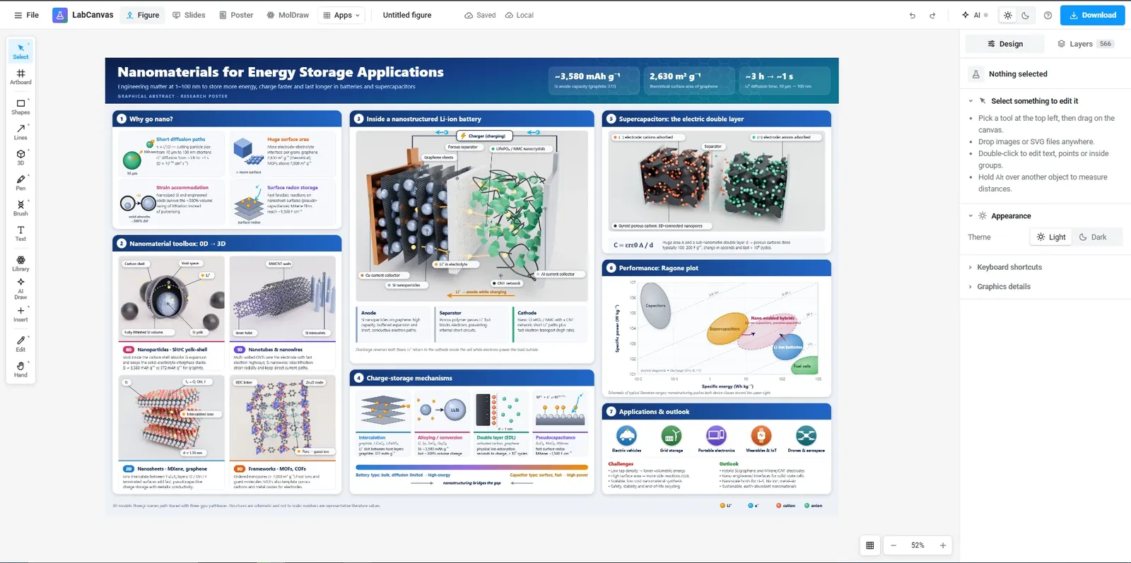
The LabCanvas Figure editor: vector scientific illustration in your browser
How to make journal figures and graphical abstracts in the LabCanvas Figure editor: vector tools, 700+ adjustable science objects, lab glassware that connects, LaTeX equations, and export at the width and DPI your journal asks for.
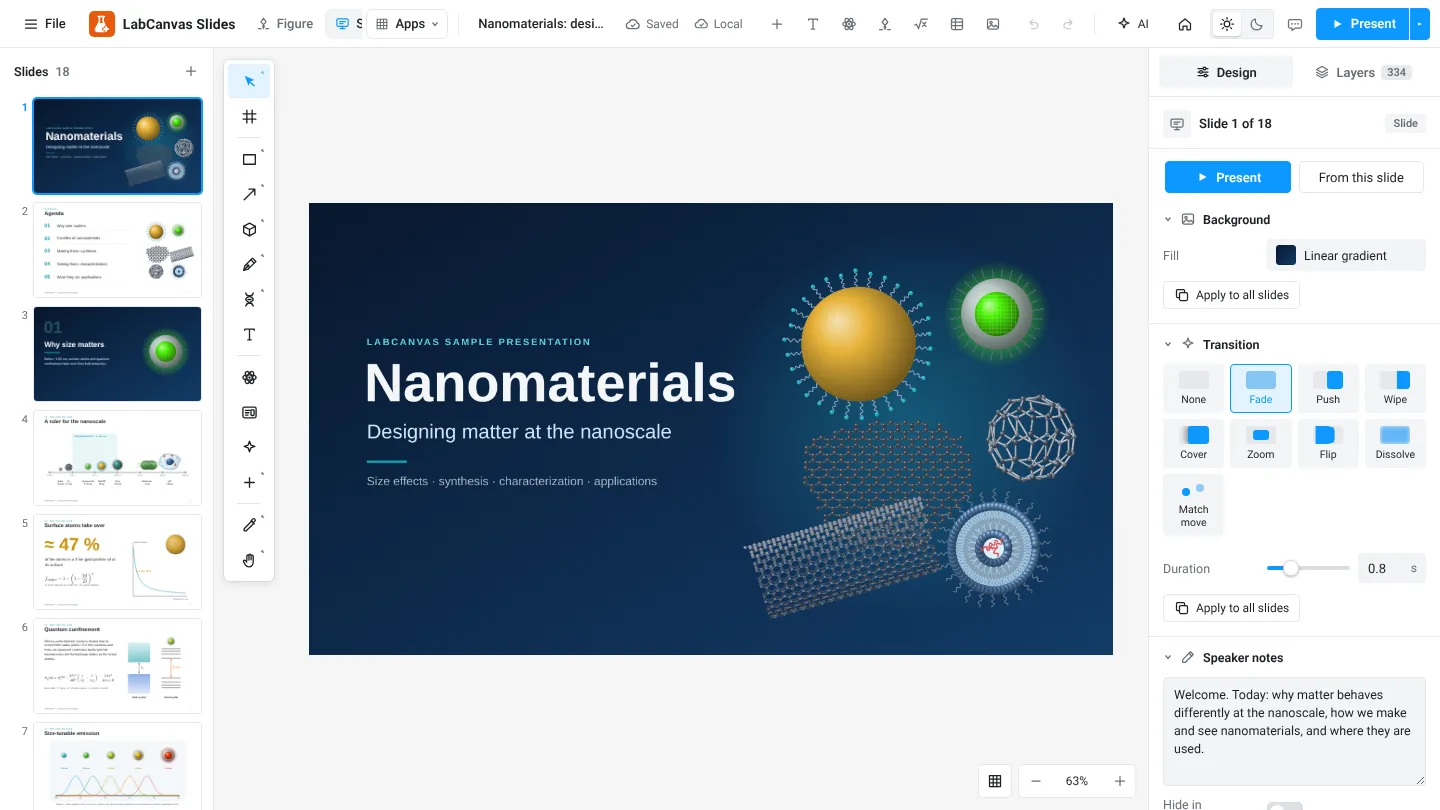
LabCanvas Slides: scientific presentations built from editable figures
How to build lab meetings, conference talks and lectures in LabCanvas Slides: a slide strip, layouts and themes, transitions and motion paths, speaker notes, full-screen presenting and a vector PDF of the deck.
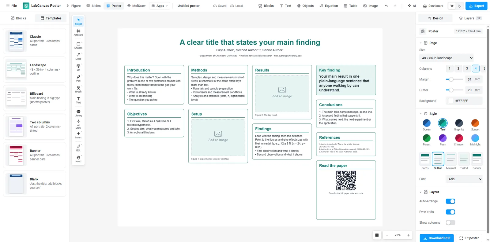
LabCanvas Research Poster: conference posters that arrange themselves
How to make an A0, A1 or 48 × 36 in research poster in LabCanvas Research Poster: blocks that stack in columns and re-flow their text, themes and block styles, QR codes, and a print-ready vector PDF.