Bacterial cell diagram with labels
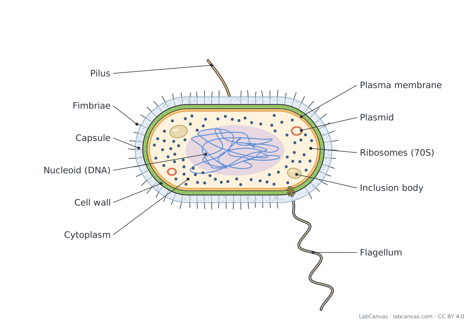
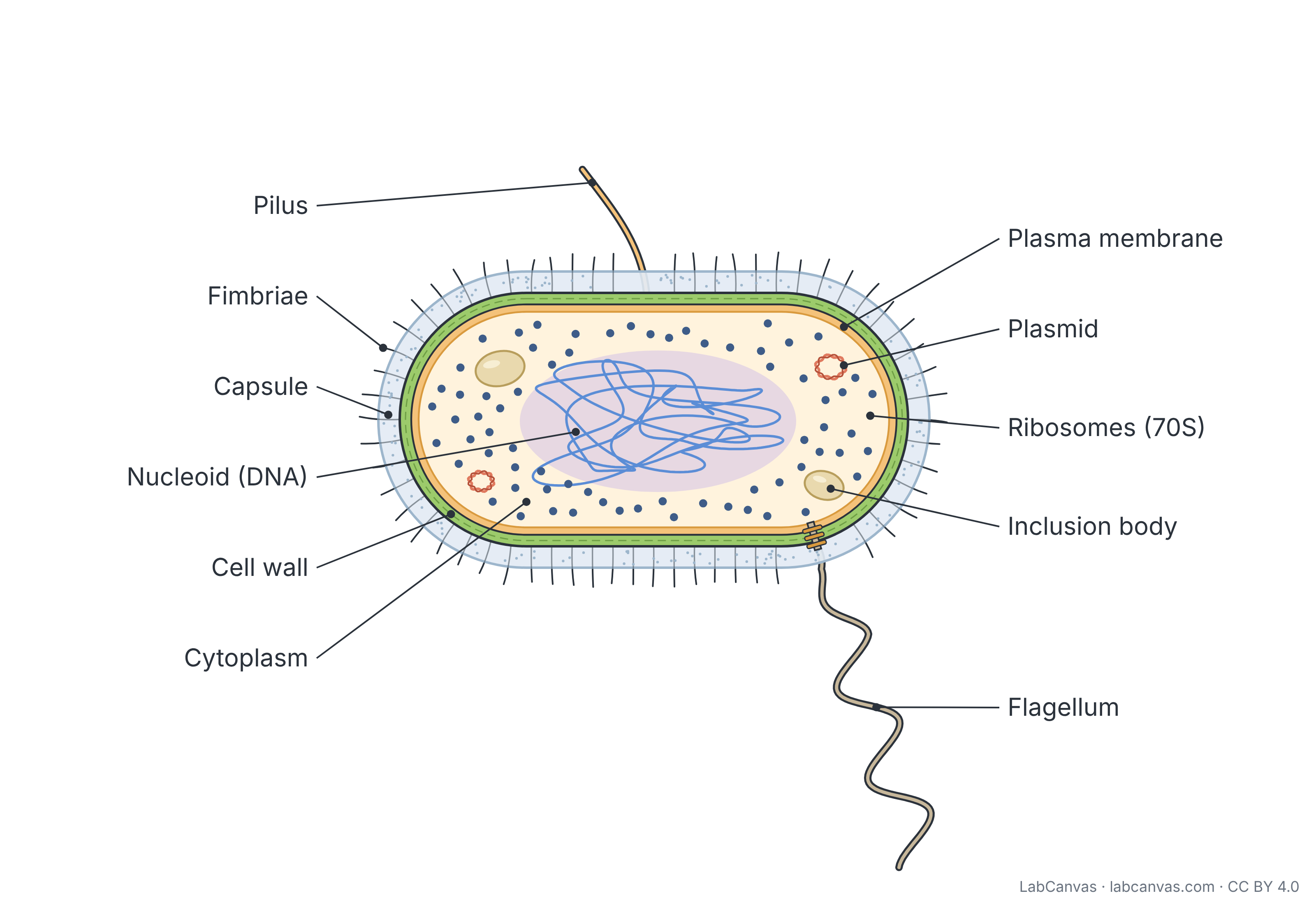
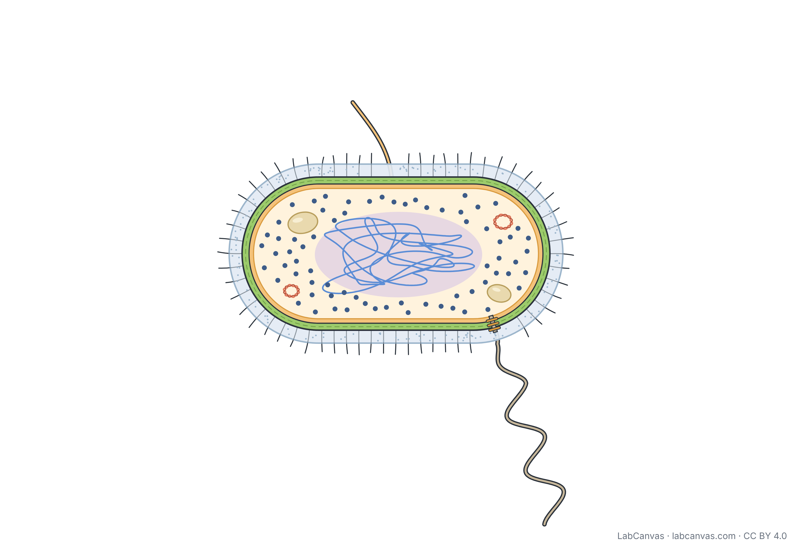
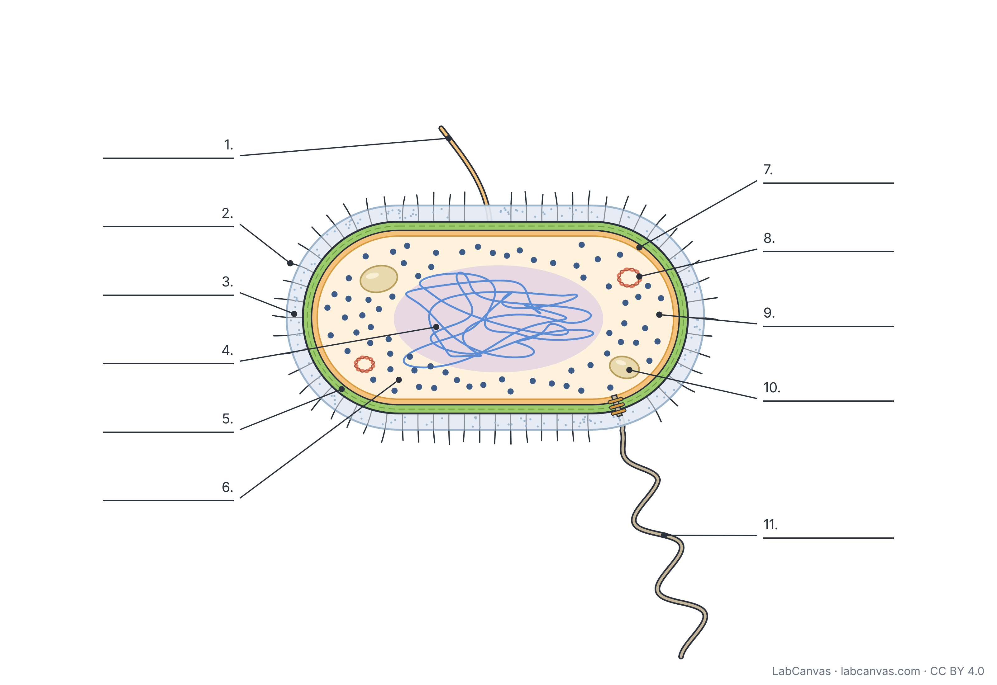
A bacterial cell is a prokaryotic cell: it has no nucleus and no membrane-bound organelles. Its DNA sits in a region of the cytoplasm called the nucleoid, often with extra small DNA circles called plasmids, and its proteins are made on 70S ribosomes. Most bacteria are wrapped in a peptidoglycan cell wall, many add a capsule, and many have flagella, pili and fimbriae. This free diagram labels 11 parts of a rod-shaped bacterium and comes unlabelled and as a numbered worksheet.
Last updated
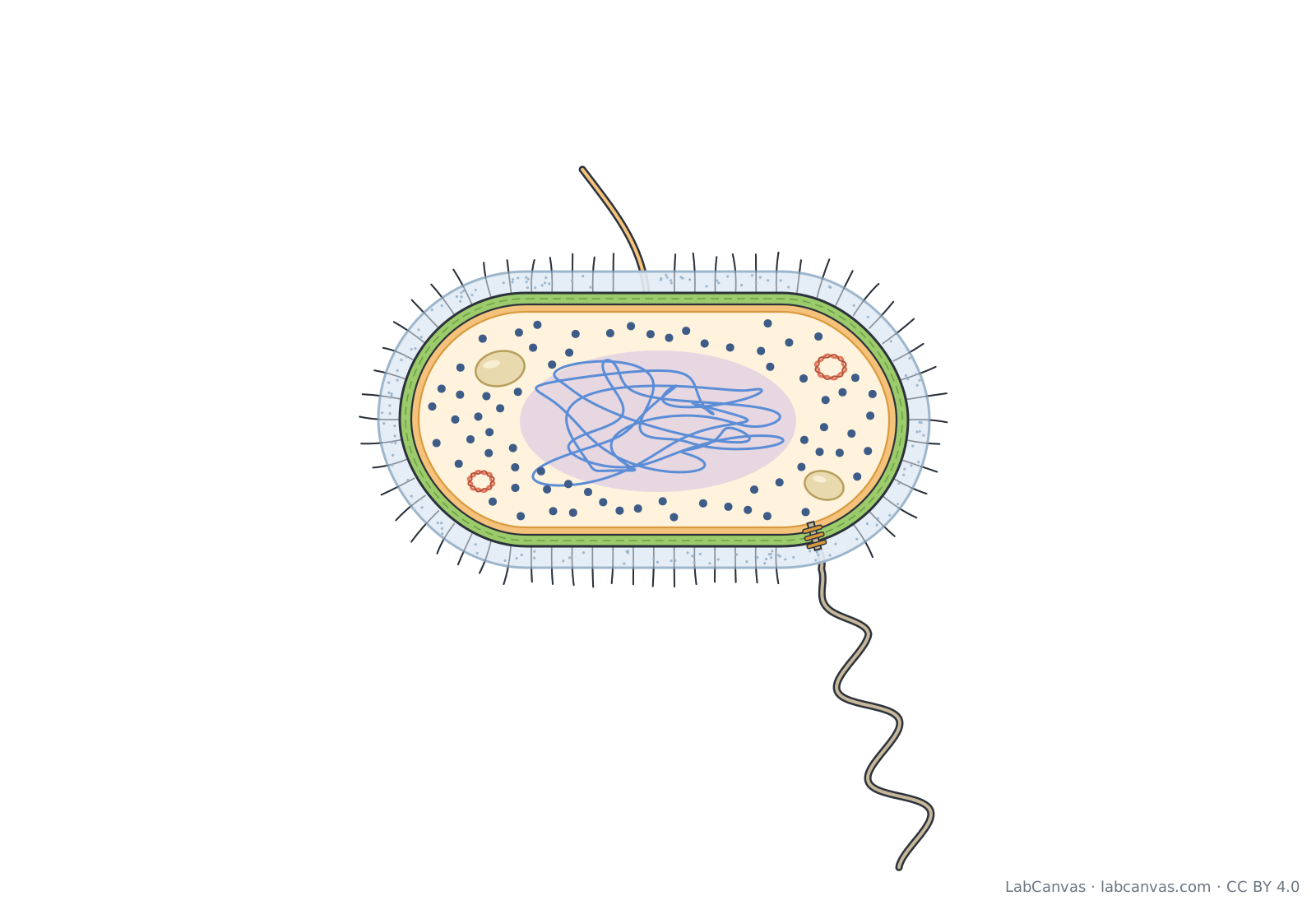
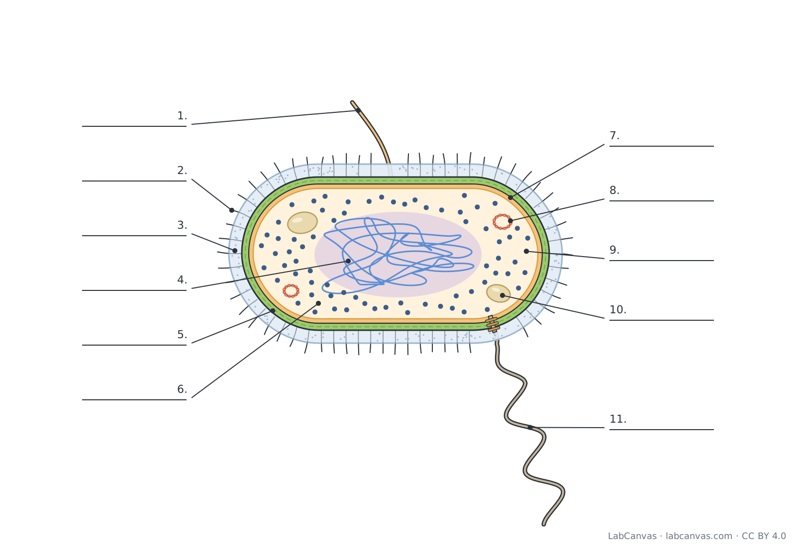
Download the bacterial cell diagram
Free to use in homework, teaching, slides, posters and publications under CC BY 4.0. Please credit: Bacterial cell diagram by LabCanvas (labcanvas.com), CC BY 4.0
Parts of the bacterial cell and what they do
| # | Part | What it is or does |
|---|---|---|
| 1 | Pilus | Long, hair-like protein appendage; a sex pilus joins two cells so DNA can pass from one to the other during conjugation. |
| 2 | Fimbriae | Many short, thin protein bristles on the surface that help the bacterium stick to surfaces and to host cells. |
| 3 | Capsule | Sticky outer layer, usually of polysaccharide, that helps the cell attach to surfaces and protects it from drying out and from being engulfed by immune cells. |
| 4 | Nucleoid (DNA) | Region of the cytoplasm, with no membrane around it, that holds the cell's chromosome: usually a single circular DNA molecule. |
| 5 | Cell wall | Rigid layer outside the plasma membrane, made of peptidoglycan in bacteria, that keeps the cell's shape and stops it bursting from osmotic pressure. |
| 6 | Cytoplasm | Gel-like contents of the cell inside the plasma membrane: water, enzymes, nutrients, ribosomes and the nucleoid. |
| 7 | Plasma membrane | Phospholipid bilayer with proteins that controls what enters and leaves the cell; in bacteria it is also where cellular respiration and other energy reactions take place. |
| 8 | Plasmid | Small circle of extra DNA, separate from the chromosome, that copies itself and often carries genes such as antibiotic resistance. |
| 9 | Ribosomes (70S) | Small particles of RNA and protein that build proteins; bacterial ribosomes are 70S, smaller than the 80S ribosomes in eukaryotic cytoplasm. |
| 10 | Inclusion body | Granule in the cytoplasm that stores reserve materials, such as glycogen, phosphate or sulfur, for later use. |
| 11 | Flagellum | Long whip-like filament anchored in the cell envelope by a basal body; it rotates like a propeller to move the cell. |
How to label a bacterial cell diagram
- Work from the outside inLabel the hazy outer coat as the capsule, the thick green layer under it as the cell wall, and the thin layer pressed against the inside of the wall as the plasma membrane.
- Label the appendagesThe long whip trailing from one end is the flagellum. The single long, thick tube is a pilus. The many short bristles all round the cell are fimbriae.
- Find the DNAThe tangled strand in the middle is the chromosome; the region it fills is the nucleoid. Note that it has no membrane around it. The small separate rings are plasmids.
- Finish the insideMark the dots as 70S ribosomes, the larger oval granules as inclusion bodies, and the fluid that fills the cell as the cytoplasm. Check your answers against the key.
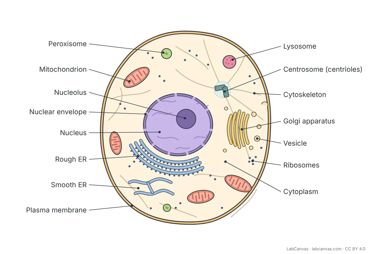
Prokaryotic (bacterial) cell vs eukaryotic cell
| Feature | Bacterial cell (prokaryote) | Eukaryotic cell (animal, plant, fungus) |
|---|---|---|
| Nucleus | No; DNA lies in the nucleoid | Yes, surrounded by a nuclear envelope |
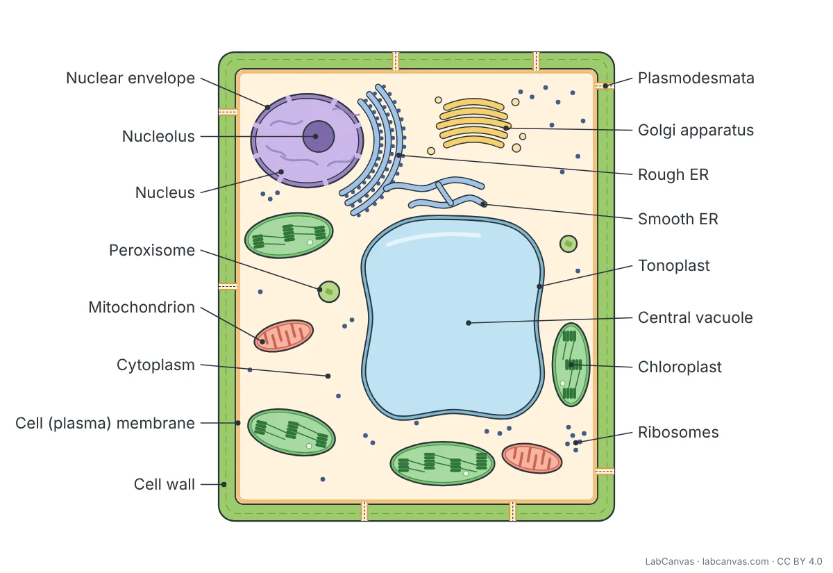
| Membrane-bound organelles | None (no mitochondria, ER or Golgi) | Many, e.g. mitochondria, ER, Golgi apparatus |
| Chromosome | Usually one circular chromosome, often plus plasmids | Several linear chromosomes in the nucleus |
| Ribosomes | 70S | 80S in the cytoplasm |
| Cell wall | Most have one, made of peptidoglycan | Plants (cellulose) and fungi (chitin) have one; animal cells do not |
| Size | Small: about 0.1 to 5.0 micrometres across | Usually much larger: about 10 to 100 micrometres |
| Plasma membrane, cytoplasm, DNA and ribosomes | Yes | Yes |
The bacterial cell envelope
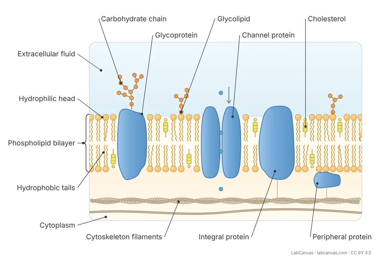
The layers around a bacterium are called the cell envelope. The innermost is the plasma membrane, a phospholipid bilayer like the membrane of every cell. Outside it, most bacteria have a cell wall of peptidoglycan, a mesh of sugars cross-linked by short chains of amino acids, which keeps the cell's shape and stops it bursting when water flows in.
In Gram-positive bacteria the peptidoglycan layer is thick. Gram-negative bacteria have a thin peptidoglycan layer with an extra outer membrane outside it, which is why the two groups stain differently in the Gram stain. Many bacteria add a capsule of polysaccharide on top, which helps them stick to surfaces and resist being engulfed by immune cells.
Flagella, pili and fimbriae
Bacterial appendages are made of protein. A flagellum is a long helical filament that rotates, driven by a motor at its base, to push the cell through liquid; a bacterium may have one or many. Fimbriae are numerous short bristles used for attachment. Pili are longer and fewer; a sex pilus draws two cells together so that DNA, often a plasmid, can be transferred in conjugation.
Where bacteria keep their DNA
Bacteria have no nucleus. Their main chromosome, usually a single circular DNA molecule, is folded into the nucleoid, a region of the cytoplasm with no membrane around it. Many bacteria also carry plasmids, small circles of DNA that replicate on their own. Plasmid genes are not usually needed for survival but can help, for example by giving resistance to an antibiotic, and plasmids can pass between cells.
Quick quiz
1. Where is the DNA of a bacterial cell found?
- In the nucleus
- In the nucleoid
- In the mitochondria
- In the capsule
Answer: b) In the nucleoid. Bacteria have no nucleus; their chromosome lies in a region of the cytoplasm called the nucleoid.
2. What is the bacterial cell wall mainly made of?
- Cellulose
- Chitin
- Peptidoglycan
- Phospholipids
Answer: c) Peptidoglycan. Most bacterial cell walls are made of peptidoglycan.
3. Which structure lets a bacterium swim?
- Flagellum
- Capsule
- Fimbriae
- Plasmid
Answer: a) Flagellum. The flagellum rotates like a propeller to move the cell.
4. What size are bacterial ribosomes?
- 80S
- 60S
- 70S
- 100S
Answer: c) 70S. Prokaryotic ribosomes are 70S; eukaryotic cytoplasmic ribosomes are 80S.
5. Which structure is used to transfer DNA between bacteria in conjugation?
- Capsule
- Sex pilus
- Inclusion body
- Cell wall
Answer: b) Sex pilus. A sex pilus joins two cells so DNA, often a plasmid, can pass between them.
Frequently asked questions
What are the parts of a bacterial cell?
A typical bacterial cell has a plasma membrane, cytoplasm, ribosomes and a nucleoid holding its DNA. Most also have a cell wall, and many have a capsule, plasmids, inclusion bodies, flagella, pili and fimbriae.
Does a bacterial cell have a nucleus?
No. Bacteria are prokaryotes, so they have no nucleus. Their DNA lies in a region of the cytoplasm called the nucleoid, which has no membrane around it.
What is the difference between a prokaryotic and a eukaryotic cell?
A prokaryotic cell, such as a bacterium, has no nucleus and no membrane-bound organelles, and it is usually much smaller. A eukaryotic cell has a nucleus and many membrane-bound organelles such as mitochondria and the Golgi apparatus.
What is the bacterial cell wall made of?
Most bacterial cell walls are made of peptidoglycan. Gram-positive bacteria have a thick peptidoglycan layer; Gram-negative bacteria have a thin one with an outer membrane outside it.
What is the difference between pili and fimbriae?
Both are protein appendages. Fimbriae are short and numerous and help the cell attach to surfaces. Pili are longer and fewer; a sex pilus is used to transfer DNA between bacteria during conjugation.
What is a plasmid in a bacterial cell?
A plasmid is a small circle of DNA separate from the main chromosome. It copies itself and often carries useful genes, such as genes for antibiotic resistance, that can be passed to other bacteria.
Do bacteria have ribosomes?
Yes. All cells have ribosomes. Bacterial ribosomes are 70S, smaller than the 80S ribosomes in the cytoplasm of eukaryotic cells, a difference some antibiotics exploit.
Sources
- OpenStax Biology 2e, 4.2 Prokaryotic Cells (CC BY 4.0)
- OpenStax Microbiology, 3.3 Unique Characteristics of Prokaryotic Cells (CC BY 4.0)
- OpenStax Biology 2e, 22.2 Structure of Prokaryotes: Bacteria and Archaea (CC BY 4.0)
- OpenStax Biology 2e, 4.3 Eukaryotic Cells (CC BY 4.0)
Facts on this page follow these introductory textbooks (OpenStax, CC BY 4.0). The diagram is a simplified illustration; organelles are not drawn to scale.
More free diagrams
Make your own science diagrams
LabCanvas is a browser workspace for scientific figures, slides and posters, with adjustable cells, organelles and molecules.
Edit this diagram