Graphical abstract template · Graphical abstracts · Graphical abstract
How the zebrafish heart rebuilds itself
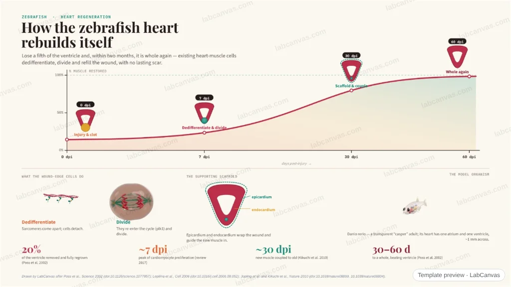
A recovery-curve graphical abstract: after losing part of its ventricle, the zebrafish heart climbs back to whole muscle over about two months. Wound-edge cardiomyocytes dedifferentiate and divide, the epicardium and endocardium build a regenerative scaffold, and the new muscle becomes electrically coupled — with no permanent scar.
Updated 6 cited papers87 editable layers
At a glance
- Category
- Graphical abstracts
- Method
- Graphical abstract
- Starting materials
- A ventricle that has lost ~20% of its muscle to resection (or ~25% to cryoinjury)
- Product
- A fully regenerated, electrically coupled ventricle within about two months
- Key result
- 20% of the ventricle resected and fully regenerated in ~2 months (Poss et al. 2002); cardiomyocyte proliferation peaks ~7 dpi, electrical coupling by ~30 dpi (Kikuchi et al. 2010; review 2017)
- Figure
- 1920 × 1080 px (16:9), 87 editable layers
Step by step
- Day 0 · injury and clotResection of ~20% of the ventricle bleeds briefly, then a fibrin clot seals the wound (Poss et al. 2002); cryoinjury (~25%) instead forms a transient fibrotic scar (González-Rosa et al. 2011)
- ~Day 7 · dedifferentiate and divideWound-edge cardiomyocytes disassemble their sarcomeres, detach and re-enter the cell cycle (plk1-dependent); gata4+ subepicardial cells contribute most of the new muscle (Jopling et al. 2010; Kikuchi et al. 2010). Proliferation peaks ~7 dpi (review 2017)
- Scaffold · epicardium & endocardiumThe epicardium expands over the wound, undergoes EMT and builds new vessels (Fgf-dependent); the endocardium proliferates (peak 3–5 dpi) — together a regenerative scaffold (Lepilina et al. 2006; review 2017)
- 30–60 days · whole and coupledNew muscle fills the wound and becomes electrically coupled to the old between 2 and 4 weeks (Kikuchi et al. 2010); the ventricle is restored in 30–60 dpi with no permanent scar (Poss et al. 2002)
Uses
- Graphical abstracts on cardiac regeneration and the zebrafish model
- Talks on cardiomyocyte dedifferentiation and proliferation
- Teaching why fish hearts regenerate and mammalian hearts scar
Papers this template follows
- Poss, Wilson & Keating, “Heart regeneration in zebrafish”, Science 2002. doi:10.1126/science.1077857method origin
- Lepilina et al., “A dynamic epicardial injury response supports progenitor cell activity during zebrafish heart regeneration”, Cell 2006. doi:10.1016/j.cell.2006.08.052primary
- Jopling et al., “Zebrafish heart regeneration occurs by cardiomyocyte dedifferentiation and proliferation”, Nature 2010. doi:10.1038/nature08899primary
- Kikuchi et al., “Primary contribution to zebrafish heart regeneration by gata4+ cardiomyocytes”, Nature 2010. doi:10.1038/nature08804primary
- González-Rosa et al., “Extensive scar formation and regression during heart regeneration after cryoinjury in zebrafish”, Development 2011. doi:10.1242/dev.060897primary
- González-Rosa, Burns & Burns, “Zebrafish heart regeneration: 15 years of discoveries”, Regeneration 2017. doi:10.1002/reg2.83review
Drawn by LabCanvas after Poss et al., Science 2002 (doi:10.1126/science.1077857); Lepilina et al., Cell 2006 (doi:10.1016/j.cell.2006.08.052); Jopling et al. and Kikuchi et al., Nature 2010 (doi:10.1038/nature08899, 10.1038/nature08804).
How templates are written and checked: editorial guidelines.
Questions
What does the “How the zebrafish heart rebuilds itself” template show?
A recovery-curve graphical abstract: after losing part of its ventricle, the zebrafish heart climbs back to whole muscle over about two months. Wound-edge cardiomyocytes dedifferentiate and divide, the epicardium and endocardium build a regenerative scaffold, and the new muscle becomes electrically coupled — with no permanent scar.
What are the steps shown in “How the zebrafish heart rebuilds itself”?
1. Day 0 · injury and clot: Resection of ~20% of the ventricle bleeds briefly, then a fibrin clot seals the wound (Poss et al. 2002); cryoinjury (~25%) instead forms a transient fibrotic scar (González-Rosa et al. 2011). 2. ~Day 7 · dedifferentiate and divide: Wound-edge cardiomyocytes disassemble their sarcomeres, detach and re-enter the cell cycle (plk1-dependent); gata4+ subepicardial cells contribute most of the new muscle (Jopling et al. 2010; Kikuchi et al. 2010). Proliferation peaks ~7 dpi (review 2017). 3. Scaffold · epicardium & endocardium: The epicardium expands over the wound, undergoes EMT and builds new vessels (Fgf-dependent); the endocardium proliferates (peak 3–5 dpi) — together a regenerative scaffold (Lepilina et al. 2006; review 2017). 4. 30–60 days · whole and coupled: New muscle fills the wound and becomes electrically coupled to the old between 2 and 4 weeks (Kikuchi et al. 2010); the ventricle is restored in 30–60 dpi with no permanent scar (Poss et al. 2002).
What are the starting materials and the product?
Starting materials: A ventricle that has lost ~20% of its muscle to resection (or ~25% to cryoinjury). Product: A fully regenerated, electrically coupled ventricle within about two months.
Which papers is “How the zebrafish heart rebuilds itself” based on?
Poss, Wilson & Keating, “Heart regeneration in zebrafish”, Science 2002 (doi:10.1126/science.1077857); Lepilina et al., “A dynamic epicardial injury response supports progenitor cell activity during zebrafish heart regeneration”, Cell 2006 (doi:10.1016/j.cell.2006.08.052); Jopling et al., “Zebrafish heart regeneration occurs by cardiomyocyte dedifferentiation and proliferation”, Nature 2010 (doi:10.1038/nature08899); Kikuchi et al., “Primary contribution to zebrafish heart regeneration by gata4+ cardiomyocytes”, Nature 2010 (doi:10.1038/nature08804); González-Rosa et al., “Extensive scar formation and regression during heart regeneration after cryoinjury in zebrafish”, Development 2011 (doi:10.1242/dev.060897); González-Rosa, Burns & Burns, “Zebrafish heart regeneration: 15 years of discoveries”, Regeneration 2017 (doi:10.1002/reg2.83).
Can I edit the “How the zebrafish heart rebuilds itself” template?
Yes. Open it in the LabCanvas editor: all 87 layers are editable. Change the text, colours and layout, then export PNG, TIFF, SVG or PDF.
Do I need an account to use this template?
Not to view it here. To open and edit it, sign in to LabCanvas or create a free account (the free plan keeps up to 3 designs). Downloading the figure is part of LabCanvas Pro (US$19 a month or US$199 a year).