Graphical abstract template · Graphical abstracts · Graphical abstract
Quantum sensing with NV centres: one defect in diamond, read by light
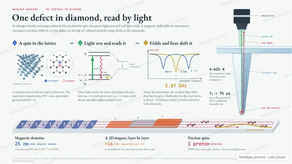
A nitrogen–vacancy centre in diamond holds an electron spin that green light sets and red fluorescence reads. A magnetic field splits its 2.87 GHz microwave resonance and heat shifts it, so a single defect at the apex of a scanning diamond tip maps magnetic domains, 2D magnets and nuclear spins at the nanoscale.
Updated 7 cited papers237 editable layers
At a glance
- Category
- Graphical abstracts
- Method
- Graphical abstract
- Starting materials
- Diamond containing nitrogen–vacancy (NV⁻) centres
- Product
- A room-temperature quantum sensor of magnetic fields and temperature, scanned at the nanoscale
- Key result
- D ≈ 2870 MHz (Sotoma et al. 2022); −74 kHz K⁻¹, ~1 ms coherence, 4–625 K (Aslam et al. 2023); 25 nm domains, 56 nT Hz⁻¹ᐟ², T₂ ≈ 75 µs in a nanopillar tip (Maletinsky et al. 2012); ≈16 µB nm⁻² in monolayer CrI₃ (Thiel et al. 2019); 1.8 mK (Kucsko et al. 2013)
- Figure
- 1920 × 1080 px (16:9), 237 editable layers
Step by step
- A spin in the latticeA nitrogen atom beside a vacancy; NV⁻ has S = 1 and a spin-triplet ground state, mₛ = 0 and ±1 split by about 2870 MHz (Sotoma et al. 2022; Mahtab et al. 2023)
- Light sets and reads itGreen light excites the centre; it fluoresces (zero-phonon line 637 nm), and microwaves that drive mₛ = 0 → ±1 dim the fluorescence through spin-dependent intersystem crossing (Sotoma et al. 2022); readout works down to a single NV (Aslam et al. 2023)
- Fields split it, heat shifts itA field lifts the ±1 degeneracy and splits the ODMR dip (Sotoma et al. 2022); the zero-field splitting moves by about −74 kHz per kelvin (Aslam et al. 2023); 1.8 mK resolved in bulk diamond (Kucsko et al. 2013)
- Scanned on a tipA single NV at the end of a diamond nanopillar AFM tip images 25 nm magnetic domains with 56 nT Hz⁻¹ᐟ² (Maletinsky et al. 2012), maps CrI₃ layer by layer (Thiel et al. 2019) and, with NMR schemes, detects down to a single proton (Aslam et al. 2023)
Uses
- Graphical abstracts on NV magnetometry and quantum sensing
- Talks introducing ODMR and scanning NV microscopy
- Teaching spin defects in wide-gap solids
Papers this template follows
- Sotoma et al., “Quantum nanodiamonds for sensing of biological quantities: Angle, temperature, and thermal conductivity”, Biophys. Physicobiol. 2022. doi:10.2142/biophysico.bppb-v19.0034review
- Aslam et al., “Quantum sensors for biomedical applications”, Nat. Rev. Phys. 2023. doi:10.1038/s42254-023-00558-3review
- Maletinsky et al., “A robust scanning diamond sensor for nanoscale imaging with single nitrogen-vacancy centres”, Nat. Nanotechnol. 2012. doi:10.1038/nnano.2012.50primary
- Thiel et al., “Probing magnetism in 2D materials at the nanoscale with single-spin microscopy”, Science 2019. doi:10.1126/science.aav6926primary
- Kucsko et al., “Nanometre-scale thermometry in a living cell”, Nature 2013. doi:10.1038/nature12373primary
- Maze et al., “Nanoscale magnetic sensing with an individual electronic spin in diamond”, Nature 2008. doi:10.1038/nature07279primary
- Mahtab et al., “High efficiency radio frequency antennas for amplifier free quantum sensing applications”, Rev. Sci. Instrum. 2023. doi:10.1063/5.0136233further reading
Drawn by LabCanvas after Sotoma et al., Biophys. Physicobiol. 2022 (doi:10.2142/biophysico.bppb-v19.0034); Aslam et al., Nat. Rev. Phys. 2023 (doi:10.1038/s42254-023-00558-3); Maletinsky et al., Nat. Nanotechnol. 2012 (doi:10.1038/nnano.2012.50); Thiel et al., Science 2019 (doi:10.1126/science.aav6926); Kucsko et al., Nature 2013 (doi:10.1038/nature12373).
How templates are written and checked: editorial guidelines.
Questions
What does the “Quantum sensing with NV centres: one defect in diamond, read by light” template show?
A nitrogen–vacancy centre in diamond holds an electron spin that green light sets and red fluorescence reads. A magnetic field splits its 2.87 GHz microwave resonance and heat shifts it, so a single defect at the apex of a scanning diamond tip maps magnetic domains, 2D magnets and nuclear spins at the nanoscale.
What are the steps shown in “Quantum sensing with NV centres: one defect in diamond, read by light”?
1. A spin in the lattice: A nitrogen atom beside a vacancy; NV⁻ has S = 1 and a spin-triplet ground state, mₛ = 0 and ±1 split by about 2870 MHz (Sotoma et al. 2022; Mahtab et al. 2023). 2. Light sets and reads it: Green light excites the centre; it fluoresces (zero-phonon line 637 nm), and microwaves that drive mₛ = 0 → ±1 dim the fluorescence through spin-dependent intersystem crossing (Sotoma et al. 2022); readout works down to a single NV (Aslam et al. 2023). 3. Fields split it, heat shifts it: A field lifts the ±1 degeneracy and splits the ODMR dip (Sotoma et al. 2022); the zero-field splitting moves by about −74 kHz per kelvin (Aslam et al. 2023); 1.8 mK resolved in bulk diamond (Kucsko et al. 2013). 4. Scanned on a tip: A single NV at the end of a diamond nanopillar AFM tip images 25 nm magnetic domains with 56 nT Hz⁻¹ᐟ² (Maletinsky et al. 2012), maps CrI₃ layer by layer (Thiel et al. 2019) and, with NMR schemes, detects down to a single proton (Aslam et al. 2023).
What are the starting materials and the product?
Starting materials: Diamond containing nitrogen–vacancy (NV⁻) centres. Product: A room-temperature quantum sensor of magnetic fields and temperature, scanned at the nanoscale.
Which papers is “Quantum sensing with NV centres: one defect in diamond, read by light” based on?
Sotoma et al., “Quantum nanodiamonds for sensing of biological quantities: Angle, temperature, and thermal conductivity”, Biophys. Physicobiol. 2022 (doi:10.2142/biophysico.bppb-v19.0034); Aslam et al., “Quantum sensors for biomedical applications”, Nat. Rev. Phys. 2023 (doi:10.1038/s42254-023-00558-3); Maletinsky et al., “A robust scanning diamond sensor for nanoscale imaging with single nitrogen-vacancy centres”, Nat. Nanotechnol. 2012 (doi:10.1038/nnano.2012.50); Thiel et al., “Probing magnetism in 2D materials at the nanoscale with single-spin microscopy”, Science 2019 (doi:10.1126/science.aav6926); Kucsko et al., “Nanometre-scale thermometry in a living cell”, Nature 2013 (doi:10.1038/nature12373); Maze et al., “Nanoscale magnetic sensing with an individual electronic spin in diamond”, Nature 2008 (doi:10.1038/nature07279); Mahtab et al., “High efficiency radio frequency antennas for amplifier free quantum sensing applications”, Rev. Sci. Instrum. 2023 (doi:10.1063/5.0136233).
Can I edit the “Quantum sensing with NV centres: one defect in diamond, read by light” template?
Yes. Open it in the LabCanvas editor: all 237 layers are editable. Change the text, colours and layout, then export PNG, TIFF, SVG or PDF.
Do I need an account to use this template?
Not to view it here. To open and edit it, sign in to LabCanvas or create a free account (the free plan keeps up to 3 designs). Downloading the figure is part of LabCanvas Pro (US$19 a month or US$199 a year).