Graphical abstract template · Nanomaterial synthesis · Thin-film hydration · Bottom-up
Liposomes by thin-film hydration and extrusion
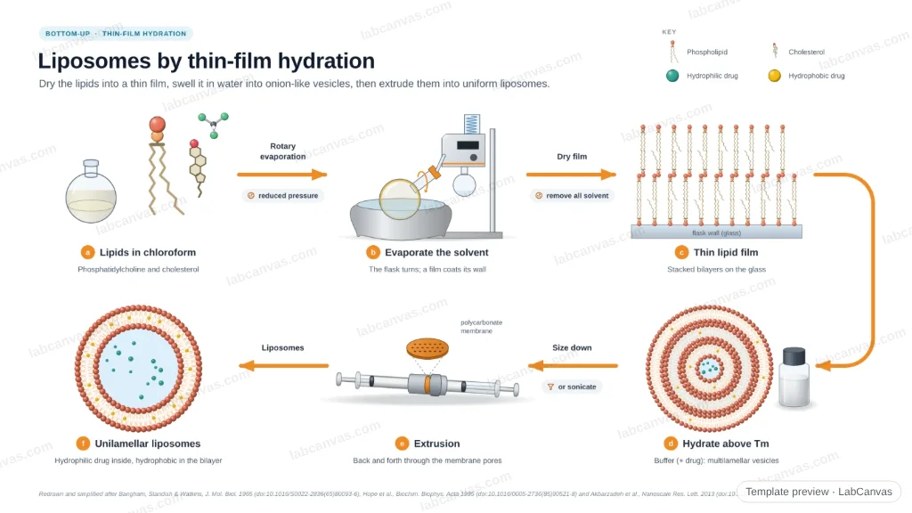
Phospholipids such as phosphatidylcholine, usually with cholesterol, are dissolved in chloroform and the solvent is removed on a rotary evaporator, leaving a thin lipid film on the flask wall. Hydrating the film with an aqueous buffer above the lipids’ phase-transition temperature swells it into multilamellar vesicles. Extrusion through a polycarbonate membrane (or sonication) turns them into unilamellar liposomes, with hydrophilic drugs in the water core and hydrophobic drugs in the bilayer.
Updated 3 cited papers68 editable layers
At a glance
- Category
- Nanomaterial synthesis
- Method
- Thin-film hydration
- Approach
- Bottom-up
- Starting materials
- Phospholipids (e.g. phosphatidylcholine) and cholesterol in chloroform; aqueous buffer (± drug)
- Product
- Unilamellar liposomes (LUVs or SUVs) carrying hydrophilic and/or hydrophobic drugs
- Typical size
- MLVs are large and polydisperse; extrusion gives vesicles close to the membrane pore size
- Key result
- Narrow size distribution after extrusion; lamellarity and loading depend on lipid composition
- Scalability
- Low to medium: lab-scale batches; extrusion and solvent removal limit scale-up
- Figure
- 1920 × 1080 px (16:9), 68 editable layers, 15 generated objects
Step by step
- Dissolve the lipidsPhospholipid and cholesterol (and a hydrophobic drug) in chloroform in a round-bottom flask
- Thin filmRemove the solvent on a rotary evaporator under reduced pressure; dry the film thoroughly
- HydrationAdd aqueous buffer (with a hydrophilic drug) above the lipid phase-transition temperature (Tm) and agitate: the film swells into multilamellar vesicles (MLVs)
- Size reductionExtrude repeatedly through a polycarbonate membrane (Hope et al.) or sonicate
- LiposomesUnilamellar vesicles; unencapsulated drug removed by dialysis or gel filtration
Strengths
- Simple and versatile; works for most lipids
- Encapsulates both hydrophilic and hydrophobic drugs
- Extrusion gives controlled, reproducible sizes
Limitations
- Uses organic solvents that must be removed completely
- Low encapsulation efficiency for hydrophilic drugs
- Hard to scale; batch-to-batch variability
Uses
- Drug delivery (e.g. anticancer drugs)
- Vaccines and adjuvants
- Model membranes
- Cosmetics and nutraceuticals
Papers this template follows
- Bangham, Standish & Watkins, “Diffusion of univalent ions across the lamellae of swollen phospholipids”, J. Mol. Biol. 1965. doi:10.1016/S0022-2836(65)80093-6method origin
- Hope et al., “Production of large unilamellar vesicles by a rapid extrusion procedure: characterization of size distribution, trapped volume and ability to maintain a membrane potential”, Biochim. Biophys. Acta 1985. doi:10.1016/0005-2736(85)90521-8primary
- Akbarzadeh et al., “Liposome: classification, preparation, and applications”, Nanoscale Res. Lett. 2013. doi:10.1186/1556-276X-8-102review
Redrawn and simplified after Bangham, Standish & Watkins, J. Mol. Biol. 1965 (doi:10.1016/S0022-2836(65)80093-6), Hope et al., Biochim. Biophys. Acta 1985 (doi:10.1016/0005-2736(85)90521-8) and Akbarzadeh et al., Nanoscale Res. Lett. 2013 (doi:10.1186/1556-276X-8-102).
How templates are written and checked: editorial guidelines.
Questions
What does the “Liposomes by thin-film hydration and extrusion” template show?
Phospholipids such as phosphatidylcholine, usually with cholesterol, are dissolved in chloroform and the solvent is removed on a rotary evaporator, leaving a thin lipid film on the flask wall. Hydrating the film with an aqueous buffer above the lipids’ phase-transition temperature swells it into multilamellar vesicles. Extrusion through a polycarbonate membrane (or sonication) turns them into unilamellar liposomes, with hydrophilic drugs in the water core and hydrophobic drugs in the bilayer.
What are the steps shown in “Liposomes by thin-film hydration and extrusion”?
1. Dissolve the lipids: Phospholipid and cholesterol (and a hydrophobic drug) in chloroform in a round-bottom flask. 2. Thin film: Remove the solvent on a rotary evaporator under reduced pressure; dry the film thoroughly. 3. Hydration: Add aqueous buffer (with a hydrophilic drug) above the lipid phase-transition temperature (Tm) and agitate: the film swells into multilamellar vesicles (MLVs). 4. Size reduction: Extrude repeatedly through a polycarbonate membrane (Hope et al.) or sonicate. 5. Liposomes: Unilamellar vesicles; unencapsulated drug removed by dialysis or gel filtration.
What are the starting materials and the product?
Starting materials: Phospholipids (e.g. phosphatidylcholine) and cholesterol in chloroform; aqueous buffer (± drug). Product: Unilamellar liposomes (LUVs or SUVs) carrying hydrophilic and/or hydrophobic drugs.
Which papers is “Liposomes by thin-film hydration and extrusion” based on?
Bangham, Standish & Watkins, “Diffusion of univalent ions across the lamellae of swollen phospholipids”, J. Mol. Biol. 1965 (doi:10.1016/S0022-2836(65)80093-6); Hope et al., “Production of large unilamellar vesicles by a rapid extrusion procedure: characterization of size distribution, trapped volume and ability to maintain a membrane potential”, Biochim. Biophys. Acta 1985 (doi:10.1016/0005-2736(85)90521-8); Akbarzadeh et al., “Liposome: classification, preparation, and applications”, Nanoscale Res. Lett. 2013 (doi:10.1186/1556-276X-8-102).
Can I edit the “Liposomes by thin-film hydration and extrusion” template?
Yes. Open it in the LabCanvas editor: all 68 layers are editable, including 15 generated objects (structures, plots, equipment) with their own settings. Change the text, colours and layout, then export PNG, TIFF, SVG or PDF.
Do I need an account to use this template?
Not to view it here. To open and edit it, sign in to LabCanvas or create a free account (the free plan keeps one design of each kind). Downloading the figure is part of LabCanvas Pro (US$19 a month or US$199 a year).