Graphical abstract template · Graphical abstracts · Graphical abstract
Carbon dots and graphene quantum dots: from synthesis to bioimaging
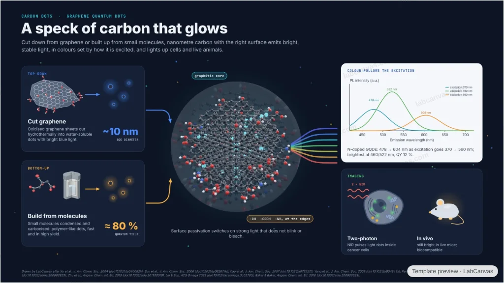
Two routes converge on one glowing dot: graphene oxide cut into ~10 nm graphene quantum dots (top-down) and small molecules carbonised into polymer-like carbon dots with quantum yields near 80 % (bottom-up). A live 3D close-up shows the graphitic core and its edge groups; to the right, excitation-dependent photoluminescence of N-doped GQDs, two-photon imaging in cells and optical imaging in live mice.
Updated 8 cited papers144 editable layers
At a glance
- Category
- Graphical abstracts
- Method
- Graphical abstract
- Starting materials
- Oxidised graphene or nanotube soot (top-down); small organic molecules (bottom-up)
- Product
- Fluorescent carbon dots and graphene quantum dots for imaging
- Key result
- QY ca. 80 % (Zhu et al. 2013); GQDs ca. 10 nm (Pan et al. 2010); emission 478–604 nm, QY 12 % (Liu & Sun 2023)
- Figure
- 1920 × 1080 px (16:9), 144 editable layers, 5 generated objects
Step by step
- Found by accidentFluorescent carbon turned up when arc-made nanotubes were purified by electrophoresis (Xu et al. 2004)
- Top-downHydrothermal cutting of oxidised graphene sheets gives water-soluble GQDs of ca. 10 nm with bright blue PL (Pan et al. 2010)
- Bottom-upA rapid, high-output route gives polymer-like carbon dots with quantum yields as high as ca. 80 % (Zhu et al. 2013)
- LightSurface passivation turns on strong, photostable PL without blinking (Sun et al. 2006); N-doped GQDs emit at 478–604 nm as the excitation moves from 370 to 560 nm, brightest at 460/522 nm, QY 12 % (Liu & Sun 2023)
- ImageTwo-photon luminescence under near-infrared pulses images cancer cells (Cao et al. 2007); the dots stay bright in live mice (Yang et al. 2009)
Uses
- Graphical abstracts on carbon dots and GQDs
- Talks on fluorescent carbon nanomaterials for imaging and sensing
- Teaching top-down and bottom-up nanomaterial synthesis
Papers this template follows
- Xu et al., “Electrophoretic analysis and purification of fluorescent single-walled carbon nanotube fragments”, J. Am. Chem. Soc. 2004. doi:10.1021/ja040082hmethod origin
- Sun et al., “Quantum-sized carbon dots for bright and colorful photoluminescence”, J. Am. Chem. Soc. 2006. doi:10.1021/ja062677dprimary
- Pan et al., “Hydrothermal route for cutting graphene sheets into blue-luminescent graphene quantum dots”, Adv. Mater. 2010. doi:10.1002/adma.200902825primary
- Zhu et al., “Highly photoluminescent carbon dots for multicolor patterning, sensors, and bioimaging”, Angew. Chem. Int. Ed. 2013. doi:10.1002/anie.201300519primary
- Cao et al., “Carbon dots for multiphoton bioimaging”, J. Am. Chem. Soc. 2007. doi:10.1021/ja073527lprimary
- Yang et al., “Carbon dots for optical imaging in vivo”, J. Am. Chem. Soc. 2009. doi:10.1021/ja904843xprimary
- Liu & Sun, “One-pot synthesis of nitrogen-doped graphene quantum dots and their applications in bioimaging and detecting copper ions in living cells”, ACS Omega 2023. doi:10.1021/acsomega.3c02705primary
- Baker & Baker, “Luminescent carbon nanodots: emergent nanolights”, Angew. Chem. Int. Ed. 2010. doi:10.1002/anie.200906623review
Drawn by LabCanvas after Xu et al., J. Am. Chem. Soc. 2004 (doi:10.1021/ja040082h); Sun et al., J. Am. Chem. Soc. 2006 (doi:10.1021/ja062677d); Cao et al., J. Am. Chem. Soc. 2007 (doi:10.1021/ja073527l); Yang et al., J. Am. Chem. Soc. 2009 (doi:10.1021/ja904843x); Pan et al., Adv. Mater. 2010 (doi:10.1002/adma.200902825); Zhu et al., Angew. Chem. Int. Ed. 2013 (doi:10.1002/anie.201300519); Liu & Sun, ACS Omega 2023 (doi:10.1021/acsomega.3c02705); Baker & Baker, Angew. Chem. Int. Ed. 2010 (doi:10.1002/anie.200906623).
How templates are written and checked: editorial guidelines.
Questions
What does the “Carbon dots and graphene quantum dots: from synthesis to bioimaging” template show?
Two routes converge on one glowing dot: graphene oxide cut into ~10 nm graphene quantum dots (top-down) and small molecules carbonised into polymer-like carbon dots with quantum yields near 80 % (bottom-up). A live 3D close-up shows the graphitic core and its edge groups; to the right, excitation-dependent photoluminescence of N-doped GQDs, two-photon imaging in cells and optical imaging in live mice.
What are the steps shown in “Carbon dots and graphene quantum dots: from synthesis to bioimaging”?
1. Found by accident: Fluorescent carbon turned up when arc-made nanotubes were purified by electrophoresis (Xu et al. 2004). 2. Top-down: Hydrothermal cutting of oxidised graphene sheets gives water-soluble GQDs of ca. 10 nm with bright blue PL (Pan et al. 2010). 3. Bottom-up: A rapid, high-output route gives polymer-like carbon dots with quantum yields as high as ca. 80 % (Zhu et al. 2013). 4. Light: Surface passivation turns on strong, photostable PL without blinking (Sun et al. 2006); N-doped GQDs emit at 478–604 nm as the excitation moves from 370 to 560 nm, brightest at 460/522 nm, QY 12 % (Liu & Sun 2023). 5. Image: Two-photon luminescence under near-infrared pulses images cancer cells (Cao et al. 2007); the dots stay bright in live mice (Yang et al. 2009).
What are the starting materials and the product?
Starting materials: Oxidised graphene or nanotube soot (top-down); small organic molecules (bottom-up). Product: Fluorescent carbon dots and graphene quantum dots for imaging.
Which papers is “Carbon dots and graphene quantum dots: from synthesis to bioimaging” based on?
Xu et al., “Electrophoretic analysis and purification of fluorescent single-walled carbon nanotube fragments”, J. Am. Chem. Soc. 2004 (doi:10.1021/ja040082h); Sun et al., “Quantum-sized carbon dots for bright and colorful photoluminescence”, J. Am. Chem. Soc. 2006 (doi:10.1021/ja062677d); Pan et al., “Hydrothermal route for cutting graphene sheets into blue-luminescent graphene quantum dots”, Adv. Mater. 2010 (doi:10.1002/adma.200902825); Zhu et al., “Highly photoluminescent carbon dots for multicolor patterning, sensors, and bioimaging”, Angew. Chem. Int. Ed. 2013 (doi:10.1002/anie.201300519); Cao et al., “Carbon dots for multiphoton bioimaging”, J. Am. Chem. Soc. 2007 (doi:10.1021/ja073527l); Yang et al., “Carbon dots for optical imaging in vivo”, J. Am. Chem. Soc. 2009 (doi:10.1021/ja904843x); Liu & Sun, “One-pot synthesis of nitrogen-doped graphene quantum dots and their applications in bioimaging and detecting copper ions in living cells”, ACS Omega 2023 (doi:10.1021/acsomega.3c02705); Baker & Baker, “Luminescent carbon nanodots: emergent nanolights”, Angew. Chem. Int. Ed. 2010 (doi:10.1002/anie.200906623).
Can I edit the “Carbon dots and graphene quantum dots: from synthesis to bioimaging” template?
Yes. Open it in the LabCanvas editor: all 144 layers are editable, including 5 generated objects (structures, plots, equipment) with their own settings. Change the text, colours and layout, then export PNG, TIFF or PDF at the width and DPI the journal asks for.
Do I need an account to use this template?
Not to view it here. To open and edit it, sign in to LabCanvas or create a free account (the free plan keeps up to 3 designs). Downloading the figure is part of LabCanvas Pro (US$19 a month or US$199 a year).