Graphical abstract template · Graphical abstracts · Graphical abstract
Cell-membrane-coated nanoparticles: red cell, platelet and cancer-cell cloaks
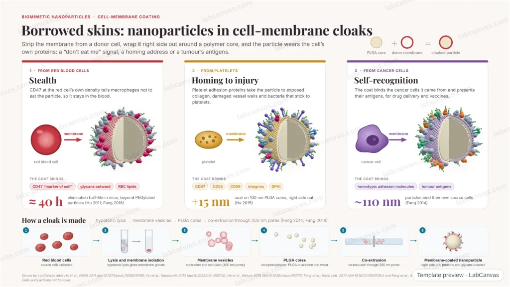
Three donor cells, three cloaks: a polymer nanoparticle wrapped right side out in the membrane of a red blood cell circulates for about 40 h, one in platelet membrane binds collagen and damaged vessels, and one in cancer-cell membrane finds its own tumour cells and carries their antigens. Live 3D particles, the proteins each coat brings and how the cloaks are made.
Updated 6 cited papers87 editable layers
At a glance
- Category
- Graphical abstracts
- Method
- Graphical abstract
- Starting materials
- Natural membranes of red blood cells, platelets or cancer cells, and PLGA nanoparticle cores
- Product
- Polymer nanoparticles wearing a donor cell’s membrane, right side out, with its proteins
- Key result
- Elimination half-life ≈ 40 h for RBC-coated particles, longer than PEGylated controls (Hu et al. 2011; Fang et al. 2018); platelet coat ≈ 15 nm on 100 nm cores (Hu et al. 2015); cancer-cell-coated particles ≈ 110 nm (Fang et al. 2014)
- Figure
- 1920 × 1080 px (16:9), 87 editable layers, 1 generated objects
Step by step
- Red blood cell cloakRBC membrane on PLGA cores: CD47 at the RBC’s density, correctly oriented (Hu et al. 2013); longer circulation than synthetic stealth coatings, still in blood at 72 h (Hu et al. 2011), an elimination half-life of about 40 h (Fang et al. 2018)
- Platelet cloakPlatelet membrane fused to 100 nm cores adds ~15 nm, carries CD47, CD55, CD59, integrins and GPVI and binds collagen and damaged vasculature (Hu et al. 2015)
- Cancer-cell cloak~110 nm particles bind their own source cells (homotypic adhesion) and present tumour antigens for vaccination (Fang et al. 2014)
- Making a cloakHypotonic lysis to membrane ghosts, vesicles by sonication and extrusion, PLGA cores, co-extrusion through 200 nm pores (Fang et al. 2014, 2018)
Uses
- Graphical abstracts on biomimetic and cell-membrane-coated nanoparticles
- Talks on stealth, homing and homotypic targeting
- Teaching how membrane coats are made
Papers this template follows
- Hu et al., “Erythrocyte membrane-camouflaged polymeric nanoparticles as a biomimetic delivery platform”, Proc. Natl. Acad. Sci. USA 2011. doi:10.1073/pnas.1106634108method origin
- Hu et al., “'Marker-of-self' functionalization of nanoscale particles through a top-down cellular membrane coating approach”, Nanoscale 2013. doi:10.1039/c3nr00015jprimary
- Luk et al., “Interfacial interactions between natural RBC membranes and synthetic polymeric nanoparticles”, Nanoscale 2014. doi:10.1039/c3nr06371bprimary
- Hu et al., “Nanoparticle biointerfacing by platelet membrane cloaking”, Nature 2015. doi:10.1038/nature15373primary
- Fang et al., “Cancer cell membrane-coated nanoparticles for anticancer vaccination and drug delivery”, Nano Lett. 2014. doi:10.1021/nl500618uprimary
- Fang, Kroll, Gao & Zhang, “Cell membrane coating nanotechnology”, Adv. Mater. 2018. doi:10.1002/adma.201706759review
Drawn by LabCanvas after Hu et al., PNAS 2011 (doi:10.1073/pnas.1106634108); Hu et al., Nanoscale 2013 (doi:10.1039/c3nr00015j); Hu et al., Nature 2015 (doi:10.1038/nature15373); Fang et al., Nano Lett. 2014 (doi:10.1021/nl500618u) and Fang et al., Adv. Mater. 2018 (doi:10.1002/adma.201706759). Cells and particles not to scale.
How templates are written and checked: editorial guidelines.
Questions
What does the “Cell-membrane-coated nanoparticles: red cell, platelet and cancer-cell cloaks” template show?
Three donor cells, three cloaks: a polymer nanoparticle wrapped right side out in the membrane of a red blood cell circulates for about 40 h, one in platelet membrane binds collagen and damaged vessels, and one in cancer-cell membrane finds its own tumour cells and carries their antigens. Live 3D particles, the proteins each coat brings and how the cloaks are made.
What are the steps shown in “Cell-membrane-coated nanoparticles: red cell, platelet and cancer-cell cloaks”?
1. Red blood cell cloak: RBC membrane on PLGA cores: CD47 at the RBC’s density, correctly oriented (Hu et al. 2013); longer circulation than synthetic stealth coatings, still in blood at 72 h (Hu et al. 2011), an elimination half-life of about 40 h (Fang et al. 2018). 2. Platelet cloak: Platelet membrane fused to 100 nm cores adds ~15 nm, carries CD47, CD55, CD59, integrins and GPVI and binds collagen and damaged vasculature (Hu et al. 2015). 3. Cancer-cell cloak: ~110 nm particles bind their own source cells (homotypic adhesion) and present tumour antigens for vaccination (Fang et al. 2014). 4. Making a cloak: Hypotonic lysis to membrane ghosts, vesicles by sonication and extrusion, PLGA cores, co-extrusion through 200 nm pores (Fang et al. 2014, 2018).
What are the starting materials and the product?
Starting materials: Natural membranes of red blood cells, platelets or cancer cells, and PLGA nanoparticle cores. Product: Polymer nanoparticles wearing a donor cell’s membrane, right side out, with its proteins.
Which papers is “Cell-membrane-coated nanoparticles: red cell, platelet and cancer-cell cloaks” based on?
Hu et al., “Erythrocyte membrane-camouflaged polymeric nanoparticles as a biomimetic delivery platform”, Proc. Natl. Acad. Sci. USA 2011 (doi:10.1073/pnas.1106634108); Hu et al., “'Marker-of-self' functionalization of nanoscale particles through a top-down cellular membrane coating approach”, Nanoscale 2013 (doi:10.1039/c3nr00015j); Luk et al., “Interfacial interactions between natural RBC membranes and synthetic polymeric nanoparticles”, Nanoscale 2014 (doi:10.1039/c3nr06371b); Hu et al., “Nanoparticle biointerfacing by platelet membrane cloaking”, Nature 2015 (doi:10.1038/nature15373); Fang et al., “Cancer cell membrane-coated nanoparticles for anticancer vaccination and drug delivery”, Nano Lett. 2014 (doi:10.1021/nl500618u); Fang, Kroll, Gao & Zhang, “Cell membrane coating nanotechnology”, Adv. Mater. 2018 (doi:10.1002/adma.201706759).
Can I edit the “Cell-membrane-coated nanoparticles: red cell, platelet and cancer-cell cloaks” template?
Yes. Open it in the LabCanvas editor: all 87 layers are editable, including 1 generated objects (structures, plots, equipment) with their own settings. Change the text, colours and layout, then export PNG, TIFF or PDF at the width and DPI the journal asks for.
Do I need an account to use this template?
Not to view it here. To open and edit it, sign in to LabCanvas or create a free account (the free plan keeps up to 3 designs). Downloading the figure is part of LabCanvas Pro (US$19 a month or US$199 a year).