Graphical abstract template · Graphical abstracts · Graphical abstract
Electrochemical biosensors: from a drop on an electrode to a number
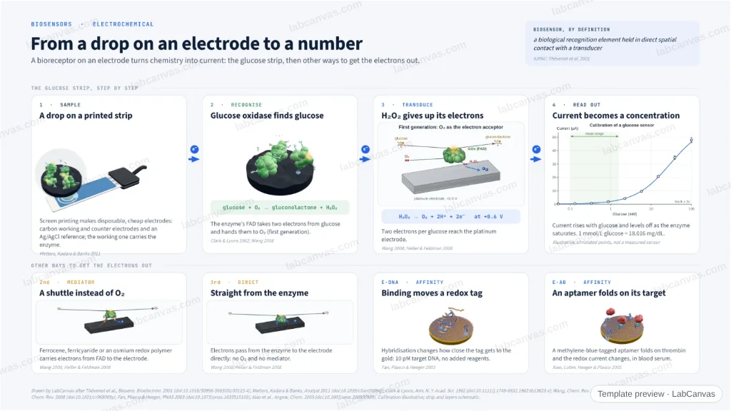
A glucose test strip followed from sample to signal: a drop on a screen-printed electrode, glucose oxidase turning glucose into gluconolactone, hydrogen peroxide oxidised at the electrode near +0.6 V, and a calibration that turns current into a concentration. Below, the other ways to get the electrons out: a redox mediator, direct electron transfer, and affinity sensors in which binding moves a redox tag (E-DNA at 10 pM, an aptamer for thrombin in serum).
Updated 7 cited papers79 editable layers
At a glance
- Category
- Graphical abstracts
- Method
- Graphical abstract
- Starting materials
- A drop of sample on a screen-printed electrode carrying a bioreceptor
- Product
- A current, calibrated into a concentration
- Key result
- H₂O₂ oxidised near +0.6 V at platinum (Wang 2008; Heller & Feldman 2008); E-DNA to 10 pM target (Fan et al. 2003); thrombin in blood serum (Xiao et al. 2005); calibration illustrative
- Figure
- 1920 × 1080 px (16:9), 79 editable layers, 3 generated objects
Step by step
- 1 · SampleA drop lands on a disposable screen-printed strip: a carbon working electrode with the enzyme, a counter electrode and an Ag/AgCl reference (Metters, Kadara & Banks 2011)
- 2 · RecogniseGlucose oxidase oxidises glucose to gluconolactone; its FAD takes the electrons and O₂ turns into H₂O₂ (first generation; Clark & Lyons 1962)
- 3 · TransduceH₂O₂ is oxidised at the electrode near +0.6 V: two electrons per glucose (Wang 2008; Heller & Feldman 2008)
- 4 · Read outA calibration turns the current into a concentration; 1 mmol/L glucose = 18.016 mg/dL
- Mediator and direct transferA mediator (ferrocene, ferricyanide, an osmium redox polymer) carries the electrons instead of O₂ (second generation), or they pass straight from the enzyme (third)
- Affinity sensorsBinding moves a redox tag: E-DNA detects 10 pM DNA (Fan et al. 2003); a methylene-blue aptamer reads thrombin in serum (Xiao et al. 2005)
Uses
- Graphical abstracts on electrochemical biosensors and glucose sensing
- Teaching how an enzyme electrode works
- Talks on point-of-care diagnostics
Papers this template follows
- Thévenot, Toth, Durst & Wilson, “Electrochemical biosensors: recommended definitions and classification”, Biosens. Bioelectron. 2001. doi:10.1016/S0956-5663(01)00115-4review
- Metters, Kadara & Banks, “New directions in screen printed electroanalytical sensors: an overview of recent developments”, Analyst 2011. doi:10.1039/c0an00894jreview
- Clark & Lyons, “Electrode systems for continuous monitoring in cardiovascular surgery”, Ann. N. Y. Acad. Sci. 1962. doi:10.1111/j.1749-6632.1962.tb13623.xprimary
- Wang, “Electrochemical glucose biosensors”, Chem. Rev. 2008. doi:10.1021/cr068123areview
- Heller & Feldman, “Electrochemical glucose sensors and their applications in diabetes management”, Chem. Rev. 2008. doi:10.1021/cr068069yreview
- Fan, Plaxco & Heeger, “Electrochemical interrogation of conformational changes as a reagentless method for the sequence-specific detection of DNA”, Proc. Natl. Acad. Sci. USA 2003. doi:10.1073/pnas.1633515100primary
- Xiao, Lubin, Heeger & Plaxco, “Label-free electronic detection of thrombin in blood serum by using an aptamer-based sensor”, Angew. Chem. Int. Ed. 2005. doi:10.1002/anie.200500989primary
Drawn by LabCanvas after Thévenot et al., Biosens. Bioelectron. 2001 (doi:10.1016/S0956-5663(01)00115-4); Metters, Kadara & Banks, Analyst 2011 (doi:10.1039/c0an00894j); Clark & Lyons, Ann. N. Y. Acad. Sci. 1962 (doi:10.1111/j.1749-6632.1962.tb13623.x); Wang, Chem. Rev. 2008 (doi:10.1021/cr068123a); Heller & Feldman, Chem. Rev. 2008 (doi:10.1021/cr068069y); Fan, Plaxco & Heeger, PNAS 2003 (doi:10.1073/pnas.1633515100); Xiao et al., Angew. Chem. 2005 (doi:10.1002/anie.200500989). Calibration illustrative; strip and layers schematic.
How templates are written and checked: editorial guidelines.
Questions
What does the “Electrochemical biosensors: from a drop on an electrode to a number” template show?
A glucose test strip followed from sample to signal: a drop on a screen-printed electrode, glucose oxidase turning glucose into gluconolactone, hydrogen peroxide oxidised at the electrode near +0.6 V, and a calibration that turns current into a concentration. Below, the other ways to get the electrons out: a redox mediator, direct electron transfer, and affinity sensors in which binding moves a redox tag (E-DNA at 10 pM, an aptamer for thrombin in serum).
What are the steps shown in “Electrochemical biosensors: from a drop on an electrode to a number”?
1. 1 · Sample: A drop lands on a disposable screen-printed strip: a carbon working electrode with the enzyme, a counter electrode and an Ag/AgCl reference (Metters, Kadara & Banks 2011). 2. 2 · Recognise: Glucose oxidase oxidises glucose to gluconolactone; its FAD takes the electrons and O₂ turns into H₂O₂ (first generation; Clark & Lyons 1962). 3. 3 · Transduce: H₂O₂ is oxidised at the electrode near +0.6 V: two electrons per glucose (Wang 2008; Heller & Feldman 2008). 4. 4 · Read out: A calibration turns the current into a concentration; 1 mmol/L glucose = 18.016 mg/dL. 5. Mediator and direct transfer: A mediator (ferrocene, ferricyanide, an osmium redox polymer) carries the electrons instead of O₂ (second generation), or they pass straight from the enzyme (third). 6. Affinity sensors: Binding moves a redox tag: E-DNA detects 10 pM DNA (Fan et al. 2003); a methylene-blue aptamer reads thrombin in serum (Xiao et al. 2005).
What are the starting materials and the product?
Starting materials: A drop of sample on a screen-printed electrode carrying a bioreceptor. Product: A current, calibrated into a concentration.
Which papers is “Electrochemical biosensors: from a drop on an electrode to a number” based on?
Thévenot, Toth, Durst & Wilson, “Electrochemical biosensors: recommended definitions and classification”, Biosens. Bioelectron. 2001 (doi:10.1016/S0956-5663(01)00115-4); Metters, Kadara & Banks, “New directions in screen printed electroanalytical sensors: an overview of recent developments”, Analyst 2011 (doi:10.1039/c0an00894j); Clark & Lyons, “Electrode systems for continuous monitoring in cardiovascular surgery”, Ann. N. Y. Acad. Sci. 1962 (doi:10.1111/j.1749-6632.1962.tb13623.x); Wang, “Electrochemical glucose biosensors”, Chem. Rev. 2008 (doi:10.1021/cr068123a); Heller & Feldman, “Electrochemical glucose sensors and their applications in diabetes management”, Chem. Rev. 2008 (doi:10.1021/cr068069y); Fan, Plaxco & Heeger, “Electrochemical interrogation of conformational changes as a reagentless method for the sequence-specific detection of DNA”, Proc. Natl. Acad. Sci. USA 2003 (doi:10.1073/pnas.1633515100); Xiao, Lubin, Heeger & Plaxco, “Label-free electronic detection of thrombin in blood serum by using an aptamer-based sensor”, Angew. Chem. Int. Ed. 2005 (doi:10.1002/anie.200500989).
Can I edit the “Electrochemical biosensors: from a drop on an electrode to a number” template?
Yes. Open it in the LabCanvas editor: all 79 layers are editable, including 3 generated objects (structures, plots, equipment) with their own settings. Change the text, colours and layout, then export PNG, TIFF or PDF at the width and DPI the journal asks for.
Do I need an account to use this template?
Not to view it here. To open and edit it, sign in to LabCanvas or create a free account (the free plan keeps up to 3 designs). Downloading the figure is part of LabCanvas Pro (US$19 a month or US$199 a year).