Graphical abstract template · Graphical abstracts · Graphical abstract
Hydrogels for tissue engineering: stiffness, relaxation, degradation and printing
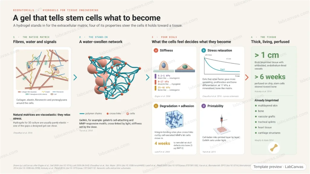
A hydrogel stands in for the extracellular matrix, and four of its properties steer the cells inside. Stiffness: soft gels push stem cells toward neurons, stiffer ones toward muscle, rigid ones toward bone. Stress relaxation: faster-relaxing gels give more spreading and a bone-like matrix. Degradability with adhesion sites: MMP-cleavable gels let cells invade and remodel a skull defect into bone in four weeks. Printability: cell-laden inks build vascularised tissues more than 1 cm thick, perfused for over six weeks.
Updated 6 cited papers98 editable layers
At a glance
- Category
- Graphical abstracts
- Method
- Graphical abstract
- Starting materials
- Cells and a hydrogel precursor (a natural or synthetic polymer with cross-linkable groups)
- Product
- A cell-laden gel that guides the cells into a tissue
- Key result
- Lineage by elasticity: 0.1–1, 8–17, 25–40 kPa (Engler et al. 2006); bone-like matrix at 17 kPa in fast-relaxing gels (Chaudhuri et al. 2016); bone in 4 weeks with 5 µg BMP-2 (Lutolf et al. 2003); > 1 cm, > 6 weeks (Kolesky et al. 2016)
- Figure
- 1920 × 1080 px (16:9), 98 editable layers, 4 generated objects
Step by step
- StiffnessMSCs on brain-like soft matrices become neurogenic, on muscle-like ones myogenic, on rigid bone-like ones osteogenic (Engler et al. 2006)
- Stress relaxationFaster-relaxing gels enhance spreading, proliferation and osteogenesis; a bone-like, mineralised matrix at 17 kPa (Chaudhuri et al. 2016)
- Degradation and adhesionIntegrin-binding sites plus MMP-cleavable cross-links let cells invade; with 5 µg BMP-2, rat cranial defects were remodelled into bone in 4 weeks (Lutolf et al. 2003)
- PrintingGelMA crosslinks under light and can be bioprinted (Yue et al. 2015); bioprinted vascularised tissues exceeded 1 cm and were perfused > 6 weeks (Kolesky et al. 2016)
Uses
- Graphical abstracts on hydrogels, biomaterials and tissue engineering
- Teaching mechanobiology and stem-cell fate
- Talks on bioprinting
Papers this template follows
- Engler, Sen, Sweeney & Discher, “Matrix elasticity directs stem cell lineage specification”, Cell 2006. doi:10.1016/j.cell.2006.06.044primary
- Chaudhuri et al., “Hydrogels with tunable stress relaxation regulate stem cell fate and activity”, Nat. Mater. 2016. doi:10.1038/nmat4489primary
- Lutolf et al., “Synthetic matrix metalloproteinase-sensitive hydrogels for the conduction of tissue regeneration: engineering cell-invasion characteristics”, Proc. Natl. Acad. Sci. USA 2003. doi:10.1073/pnas.0737381100primary
- Yue et al., “Synthesis, properties, and biomedical applications of gelatin methacryloyl (GelMA) hydrogels”, Biomaterials 2015. doi:10.1016/j.biomaterials.2015.08.045review
- Murphy & Atala, “3D bioprinting of tissues and organs”, Nat. Biotechnol. 2014. doi:10.1038/nbt.2958review
- Kolesky, Homan, Skylar-Scott & Lewis, “Three-dimensional bioprinting of thick vascularized tissues”, Proc. Natl. Acad. Sci. USA 2016. doi:10.1073/pnas.1521342113primary
Drawn by LabCanvas after Engler et al., Cell 2006 (doi:10.1016/j.cell.2006.06.044); Chaudhuri et al., Nat. Mater. 2016 (doi:10.1038/nmat4489); Lutolf et al., PNAS 2003 (doi:10.1073/pnas.0737381100); Yue et al., Biomaterials 2015 (doi:10.1016/j.biomaterials.2015.08.045); Murphy & Atala, Nat. Biotechnol. 2014 (doi:10.1038/nbt.2958); Kolesky et al., PNAS 2016 (doi:10.1073/pnas.1521342113). Network, cells and printer schematic.
How templates are written and checked: editorial guidelines.
Questions
What does the “Hydrogels for tissue engineering: stiffness, relaxation, degradation and printing” template show?
A hydrogel stands in for the extracellular matrix, and four of its properties steer the cells inside. Stiffness: soft gels push stem cells toward neurons, stiffer ones toward muscle, rigid ones toward bone. Stress relaxation: faster-relaxing gels give more spreading and a bone-like matrix. Degradability with adhesion sites: MMP-cleavable gels let cells invade and remodel a skull defect into bone in four weeks. Printability: cell-laden inks build vascularised tissues more than 1 cm thick, perfused for over six weeks.
What are the steps shown in “Hydrogels for tissue engineering: stiffness, relaxation, degradation and printing”?
1. Stiffness: MSCs on brain-like soft matrices become neurogenic, on muscle-like ones myogenic, on rigid bone-like ones osteogenic (Engler et al. 2006). 2. Stress relaxation: Faster-relaxing gels enhance spreading, proliferation and osteogenesis; a bone-like, mineralised matrix at 17 kPa (Chaudhuri et al. 2016). 3. Degradation and adhesion: Integrin-binding sites plus MMP-cleavable cross-links let cells invade; with 5 µg BMP-2, rat cranial defects were remodelled into bone in 4 weeks (Lutolf et al. 2003). 4. Printing: GelMA crosslinks under light and can be bioprinted (Yue et al. 2015); bioprinted vascularised tissues exceeded 1 cm and were perfused > 6 weeks (Kolesky et al. 2016).
What are the starting materials and the product?
Starting materials: Cells and a hydrogel precursor (a natural or synthetic polymer with cross-linkable groups). Product: A cell-laden gel that guides the cells into a tissue.
Which papers is “Hydrogels for tissue engineering: stiffness, relaxation, degradation and printing” based on?
Engler, Sen, Sweeney & Discher, “Matrix elasticity directs stem cell lineage specification”, Cell 2006 (doi:10.1016/j.cell.2006.06.044); Chaudhuri et al., “Hydrogels with tunable stress relaxation regulate stem cell fate and activity”, Nat. Mater. 2016 (doi:10.1038/nmat4489); Lutolf et al., “Synthetic matrix metalloproteinase-sensitive hydrogels for the conduction of tissue regeneration: engineering cell-invasion characteristics”, Proc. Natl. Acad. Sci. USA 2003 (doi:10.1073/pnas.0737381100); Yue et al., “Synthesis, properties, and biomedical applications of gelatin methacryloyl (GelMA) hydrogels”, Biomaterials 2015 (doi:10.1016/j.biomaterials.2015.08.045); Murphy & Atala, “3D bioprinting of tissues and organs”, Nat. Biotechnol. 2014 (doi:10.1038/nbt.2958); Kolesky, Homan, Skylar-Scott & Lewis, “Three-dimensional bioprinting of thick vascularized tissues”, Proc. Natl. Acad. Sci. USA 2016 (doi:10.1073/pnas.1521342113).
Can I edit the “Hydrogels for tissue engineering: stiffness, relaxation, degradation and printing” template?
Yes. Open it in the LabCanvas editor: all 98 layers are editable, including 4 generated objects (structures, plots, equipment) with their own settings. Change the text, colours and layout, then export PNG, TIFF or PDF at the width and DPI the journal asks for.
Do I need an account to use this template?
Not to view it here. To open and edit it, sign in to LabCanvas or create a free account (the free plan keeps up to 3 designs). Downloading the figure is part of LabCanvas Pro (US$19 a month or US$199 a year).