Graphical abstract template · Graphical abstracts · Graphical abstract
Label-free biosensors: surface plasmon resonance and graphene transistors
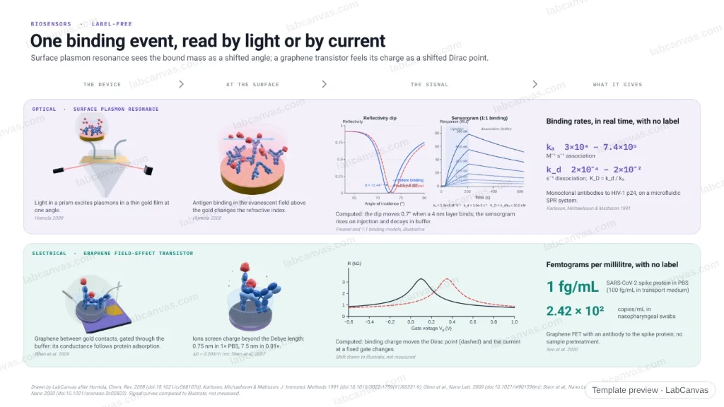
One binding event read two ways, without labels. In surface plasmon resonance, light in a prism meets a thin gold film at the resonance angle; antigen binding on the gold shifts the reflectivity dip, and the sensorgram gives association and dissociation rates in real time. In a liquid-gated graphene transistor, binding moves the Dirac point and changes the channel current; the buffer must be dilute enough that the Debye length reaches the bound charge. Numbers from the papers: kinetic constants of anti-p24 antibodies, spike protein at 1 fg/mL.
Updated 5 cited papers59 editable layers
At a glance
- Category
- Graphical abstracts
- Method
- Graphical abstract
- Starting materials
- Antibodies held on a gold film (SPR) or on graphene through pyrene linkers (GFET)
- Product
- A real-time binding curve (SPR) or a current change (GFET) when the antigen binds
- Key result
- kₐ 3 × 10⁴ – 7.4 × 10⁵ M⁻¹ s⁻¹, k_d 2 × 10⁻⁴ – 2 × 10⁻² s⁻¹ (Karlsson et al. 1991); protein at several hundred pM, transconductance ×30 in electrolyte (Ohno et al. 2009); 1 fg/mL spike protein, 2.42 × 10² copies/mL (Seo et al. 2020); signal curves computed to illustrate
- Figure
- 1920 × 1080 px (16:9), 59 editable layers
Step by step
- SPR · deviceLight in a glass prism excites surface plasmons in a thin gold film under a flow cell (Kretschmann set-up; Homola 2008)
- SPR · signalBinding on the gold changes the refractive index there and shifts the resonance angle; the sensorgram follows binding and washing in real time
- SPR · numbersAnti-p24 antibodies: kₐ 3 × 10⁴ – 7.4 × 10⁵ M⁻¹ s⁻¹, k_d 2 × 10⁻⁴ – 2 × 10⁻² s⁻¹ (Karlsson et al. 1991)
- GFET · deviceA graphene channel between gold source and drain, gated through the buffer; its conductance follows pH and protein adsorption (Ohno et al. 2009)
- GFET · Debye lengthIons screen charge beyond the Debye length, 0.75 nm in 1× PBS and 7.5 nm in 0.01× PBS, so the buffer must be controlled (Stern et al. 2007)
- GFET · numbersSpike protein at 1 fg/mL in PBS; SARS-CoV-2 at 2.42 × 10² copies/mL in swabs (Seo et al. 2020)
Uses
- Graphical abstracts on label-free and affinity biosensors
- Teaching SPR sensorgrams and binding kinetics
- Talks on graphene and FET biosensors and the Debye length
Papers this template follows
- Homola, “Surface plasmon resonance sensors for detection of chemical and biological species”, Chem. Rev. 2008. doi:10.1021/cr068107dreview
- Karlsson, Michaelsson & Mattsson, “Kinetic analysis of monoclonal antibody-antigen interactions with a new biosensor based analytical system”, J. Immunol. Methods 1991. doi:10.1016/0022-1759(91)90331-9primary
- Ohno, Maehashi, Yamashiro & Matsumoto, “Electrolyte-gated graphene field-effect transistors for detecting pH and protein adsorption”, Nano Lett. 2009. doi:10.1021/nl901596mprimary
- Stern, Wagner, Sigworth, Breaker, Fahmy & Reed, “Importance of the Debye screening length on nanowire field effect transistor sensors”, Nano Lett. 2007. doi:10.1021/nl071792zprimary
- Seo et al., “Rapid detection of COVID-19 causative virus (SARS-CoV-2) in human nasopharyngeal swab specimens using field-effect transistor-based biosensor”, ACS Nano 2020. doi:10.1021/acsnano.0c02823primary
Drawn by LabCanvas after Homola, Chem. Rev. 2008 (doi:10.1021/cr068107d); Karlsson, Michaelsson & Mattsson, J. Immunol. Methods 1991 (doi:10.1016/0022-1759(91)90331-9); Ohno et al., Nano Lett. 2009 (doi:10.1021/nl901596m); Stern et al., Nano Lett. 2007 (doi:10.1021/nl071792z); Seo et al., ACS Nano 2020 (doi:10.1021/acsnano.0c02823). Signal curves computed to illustrate, not measured.
How templates are written and checked: editorial guidelines.
Questions
What does the “Label-free biosensors: surface plasmon resonance and graphene transistors” template show?
One binding event read two ways, without labels. In surface plasmon resonance, light in a prism meets a thin gold film at the resonance angle; antigen binding on the gold shifts the reflectivity dip, and the sensorgram gives association and dissociation rates in real time. In a liquid-gated graphene transistor, binding moves the Dirac point and changes the channel current; the buffer must be dilute enough that the Debye length reaches the bound charge. Numbers from the papers: kinetic constants of anti-p24 antibodies, spike protein at 1 fg/mL.
What are the steps shown in “Label-free biosensors: surface plasmon resonance and graphene transistors”?
1. SPR · device: Light in a glass prism excites surface plasmons in a thin gold film under a flow cell (Kretschmann set-up; Homola 2008). 2. SPR · signal: Binding on the gold changes the refractive index there and shifts the resonance angle; the sensorgram follows binding and washing in real time. 3. SPR · numbers: Anti-p24 antibodies: kₐ 3 × 10⁴ – 7.4 × 10⁵ M⁻¹ s⁻¹, k_d 2 × 10⁻⁴ – 2 × 10⁻² s⁻¹ (Karlsson et al. 1991). 4. GFET · device: A graphene channel between gold source and drain, gated through the buffer; its conductance follows pH and protein adsorption (Ohno et al. 2009). 5. GFET · Debye length: Ions screen charge beyond the Debye length, 0.75 nm in 1× PBS and 7.5 nm in 0.01× PBS, so the buffer must be controlled (Stern et al. 2007). 6. GFET · numbers: Spike protein at 1 fg/mL in PBS; SARS-CoV-2 at 2.42 × 10² copies/mL in swabs (Seo et al. 2020).
What are the starting materials and the product?
Starting materials: Antibodies held on a gold film (SPR) or on graphene through pyrene linkers (GFET). Product: A real-time binding curve (SPR) or a current change (GFET) when the antigen binds.
Which papers is “Label-free biosensors: surface plasmon resonance and graphene transistors” based on?
Homola, “Surface plasmon resonance sensors for detection of chemical and biological species”, Chem. Rev. 2008 (doi:10.1021/cr068107d); Karlsson, Michaelsson & Mattsson, “Kinetic analysis of monoclonal antibody-antigen interactions with a new biosensor based analytical system”, J. Immunol. Methods 1991 (doi:10.1016/0022-1759(91)90331-9); Ohno, Maehashi, Yamashiro & Matsumoto, “Electrolyte-gated graphene field-effect transistors for detecting pH and protein adsorption”, Nano Lett. 2009 (doi:10.1021/nl901596m); Stern, Wagner, Sigworth, Breaker, Fahmy & Reed, “Importance of the Debye screening length on nanowire field effect transistor sensors”, Nano Lett. 2007 (doi:10.1021/nl071792z); Seo et al., “Rapid detection of COVID-19 causative virus (SARS-CoV-2) in human nasopharyngeal swab specimens using field-effect transistor-based biosensor”, ACS Nano 2020 (doi:10.1021/acsnano.0c02823).
Can I edit the “Label-free biosensors: surface plasmon resonance and graphene transistors” template?
Yes. Open it in the LabCanvas editor: all 59 layers are editable. Change the text, colours and layout, then export PNG, TIFF or PDF at the width and DPI the journal asks for.
Do I need an account to use this template?
Not to view it here. To open and edit it, sign in to LabCanvas or create a free account (the free plan keeps up to 3 designs). Downloading the figure is part of LabCanvas Pro (US$19 a month or US$199 a year).