Graphical abstract template · Graphical abstracts · Graphical abstract
Magnetic nanoparticles: hyperthermia, magnetic separation and MRI from one iron oxide core
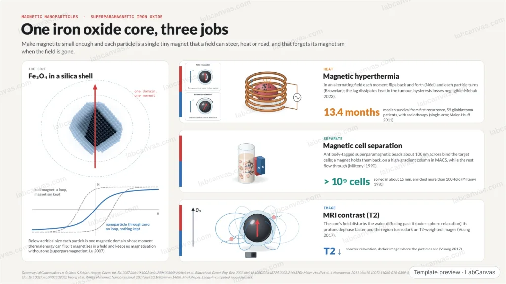
An iron oxide core small enough to be superparamagnetic magnetises in a field and forgets it when the field goes. That one property does three jobs: in an alternating field the particles heat tumours (glioblastoma patients lived a median 13.4 months after recurrence in a single-arm trial), on a high-gradient column they pull labelled cells out of a billion in 15 minutes, and in MRI they darken T2 images.
Updated 5 cited papers55 editable layers
At a glance
- Category
- Graphical abstracts
- Method
- Graphical abstract
- Starting materials
- Magnetite (Fe₃O₄) nanoparticles by co-precipitation or thermal decomposition, coated with silica or polymer
- Product
- Superparamagnetic cores for heating, separation and imaging
- Key result
- Median OS 13.4 months from first recurrence, 59 patients (Maier-Hauff et al. 2011); > 10⁹ cells in ~15 min, > 100-fold enrichment (Miltenyi et al. 1990)
- Figure
- 1920 × 1080 px (16:9), 55 editable layers, 3 generated objects
Step by step
- SuperparamagneticSmall magnetic cores magnetise in a field and keep no magnetisation without it; coatings (surfactant, polymer, silica, carbon) protect them (Lu et al. 2007)
- HeatIn an alternating field SPIONs heat by Néel and Brownian relaxation, hysteresis losses negligible (Mehak et al. 2023); with radiotherapy, recurrent glioblastoma patients had a median 13.4-month survival from recurrence (Maier-Hauff et al. 2011, single-arm)
- SeparateCells tagged with ~100 nm superparamagnetic particles are held on a high-gradient column: > 10⁹ cells in ~15 min, > 100-fold enrichment (Miltenyi et al. 1990)
- ImageThe particles’ field inhomogeneities shorten relaxation and darken T2-weighted images (outer-sphere relaxation; Vuong et al. 2017)
Uses
- Graphical abstracts on magnetic nanoparticles and theranostics
- Talks on magnetic hyperthermia, cell separation and MRI contrast
- Teaching superparamagnetism
Papers this template follows
- Lu, Salabas & Schüth, “Magnetic nanoparticles: synthesis, protection, functionalization, and application”, Angew. Chem. Int. Ed. 2007. doi:10.1002/anie.200602866review
- Mehak, Thummer & Pandey, “Surface modified iron-oxide based engineered nanomaterials for hyperthermia therapy of cancer cells”, Biotechnol. Genet. Eng. Rev. 2023. doi:10.1080/02648725.2023.2169370review
- Maier-Hauff et al., “Efficacy and safety of intratumoral thermotherapy using magnetic iron-oxide nanoparticles combined with external beam radiotherapy on patients with recurrent glioblastoma multiforme”, J. Neurooncol. 2011. doi:10.1007/s11060-010-0389-0primary
- Miltenyi, Müller, Weichel & Radbruch, “High gradient magnetic cell separation with MACS”, Cytometry 1990. doi:10.1002/cyto.990110203method origin
- Vuong, Gillis, Roch & Gossuin, “Magnetic resonance relaxation induced by superparamagnetic particles used as contrast agents in magnetic resonance imaging: a theoretical review”, WIREs Nanomed. Nanobiotechnol. 2017. doi:10.1002/wnan.1468review
Drawn by LabCanvas after Lu, Salabas & Schüth, Angew. Chem. Int. Ed. 2007 (doi:10.1002/anie.200602866); Mehak et al., Biotechnol. Genet. Eng. Rev. 2023 (doi:10.1080/02648725.2023.2169370); Maier-Hauff et al., J. Neurooncol. 2011 (doi:10.1007/s11060-010-0389-0); Miltenyi et al., Cytometry 1990 (doi:10.1002/cyto.990110203); Vuong et al., WIREs Nanomed. Nanobiotechnol. 2017 (doi:10.1002/wnan.1468). M–H shapes: Langevin computed, loop schematic.
How templates are written and checked: editorial guidelines.
Questions
What does the “Magnetic nanoparticles: hyperthermia, magnetic separation and MRI from one iron oxide core” template show?
An iron oxide core small enough to be superparamagnetic magnetises in a field and forgets it when the field goes. That one property does three jobs: in an alternating field the particles heat tumours (glioblastoma patients lived a median 13.4 months after recurrence in a single-arm trial), on a high-gradient column they pull labelled cells out of a billion in 15 minutes, and in MRI they darken T2 images.
What are the steps shown in “Magnetic nanoparticles: hyperthermia, magnetic separation and MRI from one iron oxide core”?
1. Superparamagnetic: Small magnetic cores magnetise in a field and keep no magnetisation without it; coatings (surfactant, polymer, silica, carbon) protect them (Lu et al. 2007). 2. Heat: In an alternating field SPIONs heat by Néel and Brownian relaxation, hysteresis losses negligible (Mehak et al. 2023); with radiotherapy, recurrent glioblastoma patients had a median 13.4-month survival from recurrence (Maier-Hauff et al. 2011, single-arm). 3. Separate: Cells tagged with ~100 nm superparamagnetic particles are held on a high-gradient column: > 10⁹ cells in ~15 min, > 100-fold enrichment (Miltenyi et al. 1990). 4. Image: The particles’ field inhomogeneities shorten relaxation and darken T2-weighted images (outer-sphere relaxation; Vuong et al. 2017).
What are the starting materials and the product?
Starting materials: Magnetite (Fe₃O₄) nanoparticles by co-precipitation or thermal decomposition, coated with silica or polymer. Product: Superparamagnetic cores for heating, separation and imaging.
Which papers is “Magnetic nanoparticles: hyperthermia, magnetic separation and MRI from one iron oxide core” based on?
Lu, Salabas & Schüth, “Magnetic nanoparticles: synthesis, protection, functionalization, and application”, Angew. Chem. Int. Ed. 2007 (doi:10.1002/anie.200602866); Mehak, Thummer & Pandey, “Surface modified iron-oxide based engineered nanomaterials for hyperthermia therapy of cancer cells”, Biotechnol. Genet. Eng. Rev. 2023 (doi:10.1080/02648725.2023.2169370); Maier-Hauff et al., “Efficacy and safety of intratumoral thermotherapy using magnetic iron-oxide nanoparticles combined with external beam radiotherapy on patients with recurrent glioblastoma multiforme”, J. Neurooncol. 2011 (doi:10.1007/s11060-010-0389-0); Miltenyi, Müller, Weichel & Radbruch, “High gradient magnetic cell separation with MACS”, Cytometry 1990 (doi:10.1002/cyto.990110203); Vuong, Gillis, Roch & Gossuin, “Magnetic resonance relaxation induced by superparamagnetic particles used as contrast agents in magnetic resonance imaging: a theoretical review”, WIREs Nanomed. Nanobiotechnol. 2017 (doi:10.1002/wnan.1468).
Can I edit the “Magnetic nanoparticles: hyperthermia, magnetic separation and MRI from one iron oxide core” template?
Yes. Open it in the LabCanvas editor: all 55 layers are editable, including 3 generated objects (structures, plots, equipment) with their own settings. Change the text, colours and layout, then export PNG, TIFF or PDF at the width and DPI the journal asks for.
Do I need an account to use this template?
Not to view it here. To open and edit it, sign in to LabCanvas or create a free account (the free plan keeps up to 3 designs). Downloading the figure is part of LabCanvas Pro (US$19 a month or US$199 a year).