Graphical abstract template · Graphical abstracts · Graphical abstract
The protein corona and the fate of a nanoparticle in the body
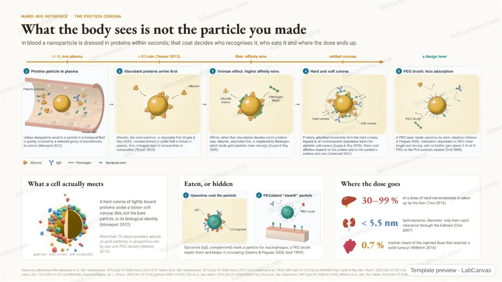
What the body sees is not the particle you made. Within half a minute of meeting plasma a nanoparticle wears a corona of proteins; higher-affinity proteins then displace the first arrivals and a hard and a soft corona settle. Opsonins hand it to macrophages unless a PEG brush hides it, and most of a dose ends in the liver: 30–99 % for hard nanomaterials, a median 0.7 % in tumours, and only particles under 5.5 nm leave through the kidneys.
Updated 11 cited papers39 editable layers
At a glance
- Category
- Graphical abstracts
- Method
- Graphical abstract
- Starting materials
- A nanoparticle entering blood plasma
- Product
- The particle’s biological identity (its corona) and where it ends up
- Key result
- Corona in < 0.5 min (Tenzer et al. 2013); liver 30–99 % (Tsoi et al. 2016); renal cut-off 5.5 nm (Choi et al. 2007); tumour 0.7 % median (Wilhelm et al. 2016)
- Figure
- 1920 × 1080 px (16:9), 39 editable layers, 5 generated objects
Step by step
- Corona in secondsPlasma coronas form in under half a minute and then change more in amount than in composition (Tenzer et al. 2013); the corona is the particle’s biological identity (Monopoli et al. 2012)
- Affinity winsProteins compete for the surface (Cedervall et al. 2007); fibrinogen displaces albumin on gold (Vroman effect); a hard, irreversible corona under a soft, exchangeable one (Gupta & Roy 2020)
- Eaten or hiddenOpsonins mark particles for MPS macrophages; a PEG brush repels them and prolongs circulation (Owens & Peppas 2006; Gref et al. 1994)
- Where the dose goesLiver 30–99 % for hard nanomaterials (Tsoi et al. 2016); kidneys only below a 5.5 nm hydrodynamic diameter (Choi et al. 2007); a median 0.7 % reaches solid tumours (Wilhelm et al. 2016)
Uses
- Graphical abstracts on the nano–bio interface and the protein corona
- Nanomedicine reviews on biodistribution and clearance
- Teaching why PEGylation matters
Papers this template follows
- Monopoli et al., “Biomolecular coronas provide the biological identity of nanosized materials”, Nat. Nanotechnol. 2012. doi:10.1038/nnano.2012.207primary
- Tenzer et al., “Rapid formation of plasma protein corona critically affects nanoparticle pathophysiology”, Nat. Nanotechnol. 2013. doi:10.1038/nnano.2013.181primary
- Cedervall et al., “Understanding the nanoparticle–protein corona using methods to quantify exchange rates and affinities of proteins for nanoparticles”, Proc. Natl. Acad. Sci. USA 2007. doi:10.1073/pnas.0608582104primary
- Walkey et al., “Nanoparticle size and surface chemistry determine serum protein adsorption and macrophage uptake”, J. Am. Chem. Soc. 2012. doi:10.1021/ja2084338primary
- Gupta & Roy, “How corona formation impacts nanomaterials as drug carriers”, Mol. Pharm. 2020. doi:10.1021/acs.molpharmaceut.9b01111review
- Owens & Peppas, “Opsonization, biodistribution, and pharmacokinetics of polymeric nanoparticles”, Int. J. Pharm. 2006. doi:10.1016/j.ijpharm.2005.10.010review
- Gref et al., “Biodegradable long-circulating polymeric nanospheres”, Science 1994. doi:10.1126/science.8128245method origin
- Gref et al., “'Stealth' corona-core nanoparticles surface modified by polyethylene glycol (PEG): influences of the corona (PEG chain length and surface density) and of the core composition on phagocytic uptake and plasma protein adsorption”, Colloids Surf. B 2000. doi:10.1016/s0927-7765(99)00156-3primary
- Tsoi et al., “Mechanism of hard-nanomaterial clearance by the liver”, Nat. Mater. 2016. doi:10.1038/nmat4718primary
- Choi et al., “Renal clearance of quantum dots”, Nat. Biotechnol. 2007. doi:10.1038/nbt1340primary
- Wilhelm et al., “Analysis of nanoparticle delivery to tumours”, Nat. Rev. Mater. 2016. doi:10.1038/natrevmats.2016.14primary
Drawn by LabCanvas after Monopoli et al., Nat. Nanotechnol. 2012 (doi:10.1038/nnano.2012.207); Tenzer et al., Nat. Nanotechnol. 2013 (doi:10.1038/nnano.2013.181); Cedervall et al., PNAS 2007 (doi:10.1073/pnas.0608582104); Gupta & Roy, Mol. Pharm. 2020 (doi:10.1021/acs.molpharmaceut.9b01111); Walkey et al., J. Am. Chem. Soc. 2012 (doi:10.1021/ja2084338); Owens & Peppas, Int. J. Pharm. 2006 (doi:10.1016/j.ijpharm.2005.10.010); Tsoi et al., Nat. Mater. 2016 (doi:10.1038/nmat4718); Choi et al., Nat. Biotechnol. 2007 (doi:10.1038/nbt1340); Wilhelm et al., Nat. Rev. Mater. 2016 (doi:10.1038/natrevmats.2016.14). Schematic, not to scale.
How templates are written and checked: editorial guidelines.
Questions
What does the “The protein corona and the fate of a nanoparticle in the body” template show?
What the body sees is not the particle you made. Within half a minute of meeting plasma a nanoparticle wears a corona of proteins; higher-affinity proteins then displace the first arrivals and a hard and a soft corona settle. Opsonins hand it to macrophages unless a PEG brush hides it, and most of a dose ends in the liver: 30–99 % for hard nanomaterials, a median 0.7 % in tumours, and only particles under 5.5 nm leave through the kidneys.
What are the steps shown in “The protein corona and the fate of a nanoparticle in the body”?
1. Corona in seconds: Plasma coronas form in under half a minute and then change more in amount than in composition (Tenzer et al. 2013); the corona is the particle’s biological identity (Monopoli et al. 2012). 2. Affinity wins: Proteins compete for the surface (Cedervall et al. 2007); fibrinogen displaces albumin on gold (Vroman effect); a hard, irreversible corona under a soft, exchangeable one (Gupta & Roy 2020). 3. Eaten or hidden: Opsonins mark particles for MPS macrophages; a PEG brush repels them and prolongs circulation (Owens & Peppas 2006; Gref et al. 1994). 4. Where the dose goes: Liver 30–99 % for hard nanomaterials (Tsoi et al. 2016); kidneys only below a 5.5 nm hydrodynamic diameter (Choi et al. 2007); a median 0.7 % reaches solid tumours (Wilhelm et al. 2016).
What are the starting materials and the product?
Starting materials: A nanoparticle entering blood plasma. Product: The particle’s biological identity (its corona) and where it ends up.
Which papers is “The protein corona and the fate of a nanoparticle in the body” based on?
Monopoli et al., “Biomolecular coronas provide the biological identity of nanosized materials”, Nat. Nanotechnol. 2012 (doi:10.1038/nnano.2012.207); Tenzer et al., “Rapid formation of plasma protein corona critically affects nanoparticle pathophysiology”, Nat. Nanotechnol. 2013 (doi:10.1038/nnano.2013.181); Cedervall et al., “Understanding the nanoparticle–protein corona using methods to quantify exchange rates and affinities of proteins for nanoparticles”, Proc. Natl. Acad. Sci. USA 2007 (doi:10.1073/pnas.0608582104); Walkey et al., “Nanoparticle size and surface chemistry determine serum protein adsorption and macrophage uptake”, J. Am. Chem. Soc. 2012 (doi:10.1021/ja2084338); Gupta & Roy, “How corona formation impacts nanomaterials as drug carriers”, Mol. Pharm. 2020 (doi:10.1021/acs.molpharmaceut.9b01111); Owens & Peppas, “Opsonization, biodistribution, and pharmacokinetics of polymeric nanoparticles”, Int. J. Pharm. 2006 (doi:10.1016/j.ijpharm.2005.10.010); Gref et al., “Biodegradable long-circulating polymeric nanospheres”, Science 1994 (doi:10.1126/science.8128245); Gref et al., “'Stealth' corona-core nanoparticles surface modified by polyethylene glycol (PEG): influences of the corona (PEG chain length and surface density) and of the core composition on phagocytic uptake and plasma protein adsorption”, Colloids Surf. B 2000 (doi:10.1016/s0927-7765(99)00156-3); Tsoi et al., “Mechanism of hard-nanomaterial clearance by the liver”, Nat. Mater. 2016 (doi:10.1038/nmat4718); Choi et al., “Renal clearance of quantum dots”, Nat. Biotechnol. 2007 (doi:10.1038/nbt1340); Wilhelm et al., “Analysis of nanoparticle delivery to tumours”, Nat. Rev. Mater. 2016 (doi:10.1038/natrevmats.2016.14).
Can I edit the “The protein corona and the fate of a nanoparticle in the body” template?
Yes. Open it in the LabCanvas editor: all 39 layers are editable, including 5 generated objects (structures, plots, equipment) with their own settings. Change the text, colours and layout, then export PNG, TIFF or PDF at the width and DPI the journal asks for.
Do I need an account to use this template?
Not to view it here. To open and edit it, sign in to LabCanvas or create a free account (the free plan keeps up to 3 designs). Downloading the figure is part of LabCanvas Pro (US$19 a month or US$199 a year).