Graphical abstract template · Cancer biology · Graphical abstract
Antibody–drug conjugate mechanism
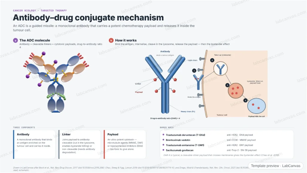
A figure of how an antibody–drug conjugate works. The 3D model shows the ADC — a monoclonal antibody carrying cytotoxic payloads on cleavable linkers at a drug-to-antibody ratio of 4 — and the 2D mechanism takes it through binding the tumour antigen, internalisation, lysosomal linker cleavage, payload release and the bystander effect. The three components and named ADCs are set out alongside.
Updated 3 cited papers45 editable layers
At a glance
- Category
- Cancer biology
- Method
- Graphical abstract
- Product
- A monoclonal antibody that delivers a cytotoxic payload inside the tumour cell
- Key result
- A typical drug-to-antibody ratio (DAR) is 4; payloads are ultra-potent microtubule agents (MMAE, DM1) or topoisomerase inhibitors (DXd) (Beck et al. 2017)
- Figure
- 1920 × 1080 px (16:9), 45 editable layers, 1 generated objects
Step by step
- Three partsA monoclonal antibody, a chemical linker (cleavable or non-cleavable) and a cytotoxic payload, at a defined drug-to-antibody ratio (Beck et al. 2017)
- Bind & internaliseThe antibody binds its tumour-cell antigen and the complex is taken up into an endosome (Beck et al. 2017)
- ReleaseIn the lysosome the linker is cleaved (or the antibody degraded), freeing the payload to kill the cell (Beck et al. 2017)
- Bystander effectMembrane-permeable payloads can diffuse out and kill neighbouring antigen-low cells (Chau et al. 2019; Drago et al. 2021)
Uses
- Targeted-therapy review and graphical-abstract figures
- Talks on ADC design
- Teaching the ADC mechanism and components
Papers this template follows
- Beck et al., “Strategies and challenges for the next generation of antibody–drug conjugates”, Nat. Rev. Drug Discov. 2017. doi:10.1038/nrd.2016.268review
- Chau, Steeg & Figg, “Antibody–drug conjugates for cancer”, Lancet 2019. doi:10.1016/S0140-6736(19)31774-Xreview
- Drago, Modi & Chandarlapaty, “Unlocking the potential of antibody–drug conjugates for cancer therapy”, Nat. Rev. Clin. Oncol. 2021. doi:10.1038/s41571-021-00470-8review
Drawn in LabCanvas after Beck et al., Nat. Rev. Drug Discov. 2017 (doi:10.1038/nrd.2016.268); Chau, Steeg & Figg, Lancet 2019 (doi:10.1016/S0140-6736(19)31774-X); and Drago, Modi & Chandarlapaty, Nat. Rev. Clin. Oncol. 2021 (doi:10.1038/s41571-021-00470-8). Domains enlarged to read; schematic.
How templates are written and checked: editorial guidelines.
Questions
What does the “Antibody–drug conjugate mechanism” template show?
A figure of how an antibody–drug conjugate works. The 3D model shows the ADC — a monoclonal antibody carrying cytotoxic payloads on cleavable linkers at a drug-to-antibody ratio of 4 — and the 2D mechanism takes it through binding the tumour antigen, internalisation, lysosomal linker cleavage, payload release and the bystander effect. The three components and named ADCs are set out alongside.
What are the steps shown in “Antibody–drug conjugate mechanism”?
1. Three parts: A monoclonal antibody, a chemical linker (cleavable or non-cleavable) and a cytotoxic payload, at a defined drug-to-antibody ratio (Beck et al. 2017). 2. Bind & internalise: The antibody binds its tumour-cell antigen and the complex is taken up into an endosome (Beck et al. 2017). 3. Release: In the lysosome the linker is cleaved (or the antibody degraded), freeing the payload to kill the cell (Beck et al. 2017). 4. Bystander effect: Membrane-permeable payloads can diffuse out and kill neighbouring antigen-low cells (Chau et al. 2019; Drago et al. 2021).
Which papers is “Antibody–drug conjugate mechanism” based on?
Beck et al., “Strategies and challenges for the next generation of antibody–drug conjugates”, Nat. Rev. Drug Discov. 2017 (doi:10.1038/nrd.2016.268); Chau, Steeg & Figg, “Antibody–drug conjugates for cancer”, Lancet 2019 (doi:10.1016/S0140-6736(19)31774-X); Drago, Modi & Chandarlapaty, “Unlocking the potential of antibody–drug conjugates for cancer therapy”, Nat. Rev. Clin. Oncol. 2021 (doi:10.1038/s41571-021-00470-8).
Can I edit the “Antibody–drug conjugate mechanism” template?
Yes. Open it in the LabCanvas editor: all 45 layers are editable, including 1 generated objects (structures, plots, equipment) with their own settings. Change the text, colours and layout, then export PNG, TIFF or PDF at the width and DPI the journal asks for.
Do I need an account to use this template?
Not to view it here. To open and edit it, sign in to LabCanvas or create a free account (the free plan keeps up to 3 designs). Downloading the figure is part of LabCanvas Pro (US$19 a month or US$199 a year).