Graphical abstract template · Cancer biology · Graphical abstract
The tumour microenvironment
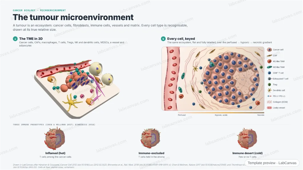
A figure of the tumour microenvironment: a 3D cross-section with every cell type recognisable — cancer cells, cancer-associated fibroblasts, M1 and M2 macrophages, CD8⁺ T cells, Tregs, NK and dendritic cells, MDSCs, endothelium, pericytes and adipocytes in their collagen matrix — beside a fully labelled 2D scene and the three immune phenotypes (inflamed, excluded, desert). Cell sizes and the oxygen diffusion limit are to scale.
Updated 4 cited papers26 editable layers
At a glance
- Category
- Cancer biology
- Method
- Graphical abstract
- Product
- The cellular ecosystem of a solid tumour: cancer cells, stroma, vasculature and immune infiltrate
- Key result
- A CD8⁺ T cell is ~7–10 µm, a macrophage ~15–20 µm; O₂ reaches ~100–200 µm from a capillary before hypoxia and necrosis (Thomlinson & Gray 1955)
- Figure
- 1920 × 1080 px (16:9), 26 editable layers, 1 generated objects
Step by step
- Cancer cells and stromaNests of cancer cells among cancer-associated fibroblasts and a remodelled collagen matrix (Hanahan & Coussens 2012)
- Immune infiltrateCD8⁺ T cells, Tregs, NK cells, dendritic cells, M1- and M2-like macrophages and MDSCs populate the tumour (Hanahan & Coussens 2012)
- Vasculature and oxygenLeaky vessels with loose pericytes; beyond ~150 µm from a vessel, tissue turns hypoxic and then necrotic (Thomlinson & Gray 1955)
- Three immune phenotypesInflamed (T cells among the cancer cells), excluded (T cells held in the stroma) and desert (few or no T cells) (Chen & Mellman 2017; Binnewies 2018)
Uses
- Review and graphical-abstract figures
- Immuno-oncology talks and posters
- Teaching the cellular ecosystem of tumours
Papers this template follows
- Hanahan & Coussens, “Accessories to the crime: functions of cells recruited to the tumor microenvironment”, Cancer Cell 2012. doi:10.1016/j.ccr.2012.02.022review
- Binnewies et al., “Understanding the tumor immune microenvironment (TIME) for effective therapy”, Nat. Med. 2018. doi:10.1038/s41591-018-0014-xreview
- Chen & Mellman, “Elements of cancer immunity and the cancer–immune set point”, Nature 2017. doi:10.1038/nature21349review
- Thomlinson & Gray, “The histological structure of some human lung cancers and the possible implications for radiotherapy”, Br. J. Cancer 1955. doi:10.1038/bjc.1955.55primary
Drawn in LabCanvas after Hanahan & Coussens, Cancer Cell 2012 (doi:10.1016/j.ccr.2012.02.022); Binnewies et al., Nat. Med. 2018 (doi:10.1038/s41591-018-0014-x); Chen & Mellman, Nature 2017 (doi:10.1038/nature21349); and Thomlinson & Gray, Br. J. Cancer 1955 (doi:10.1038/bjc.1955.55). Cells at their relative sizes; schematic.
How templates are written and checked: editorial guidelines.
Questions
What does the “The tumour microenvironment” template show?
A figure of the tumour microenvironment: a 3D cross-section with every cell type recognisable — cancer cells, cancer-associated fibroblasts, M1 and M2 macrophages, CD8⁺ T cells, Tregs, NK and dendritic cells, MDSCs, endothelium, pericytes and adipocytes in their collagen matrix — beside a fully labelled 2D scene and the three immune phenotypes (inflamed, excluded, desert). Cell sizes and the oxygen diffusion limit are to scale.
What are the steps shown in “The tumour microenvironment”?
1. Cancer cells and stroma: Nests of cancer cells among cancer-associated fibroblasts and a remodelled collagen matrix (Hanahan & Coussens 2012). 2. Immune infiltrate: CD8⁺ T cells, Tregs, NK cells, dendritic cells, M1- and M2-like macrophages and MDSCs populate the tumour (Hanahan & Coussens 2012). 3. Vasculature and oxygen: Leaky vessels with loose pericytes; beyond ~150 µm from a vessel, tissue turns hypoxic and then necrotic (Thomlinson & Gray 1955). 4. Three immune phenotypes: Inflamed (T cells among the cancer cells), excluded (T cells held in the stroma) and desert (few or no T cells) (Chen & Mellman 2017; Binnewies 2018).
Which papers is “The tumour microenvironment” based on?
Hanahan & Coussens, “Accessories to the crime: functions of cells recruited to the tumor microenvironment”, Cancer Cell 2012 (doi:10.1016/j.ccr.2012.02.022); Binnewies et al., “Understanding the tumor immune microenvironment (TIME) for effective therapy”, Nat. Med. 2018 (doi:10.1038/s41591-018-0014-x); Chen & Mellman, “Elements of cancer immunity and the cancer–immune set point”, Nature 2017 (doi:10.1038/nature21349); Thomlinson & Gray, “The histological structure of some human lung cancers and the possible implications for radiotherapy”, Br. J. Cancer 1955 (doi:10.1038/bjc.1955.55).
Can I edit the “The tumour microenvironment” template?
Yes. Open it in the LabCanvas editor: all 26 layers are editable, including 1 generated objects (structures, plots, equipment) with their own settings. Change the text, colours and layout, then export PNG, TIFF or PDF at the width and DPI the journal asks for.
Do I need an account to use this template?
Not to view it here. To open and edit it, sign in to LabCanvas or create a free account (the free plan keeps up to 3 designs). Downloading the figure is part of LabCanvas Pro (US$19 a month or US$199 a year).