Graphical abstract template · Cancer biology · Graphical abstract
Immune checkpoint blockade
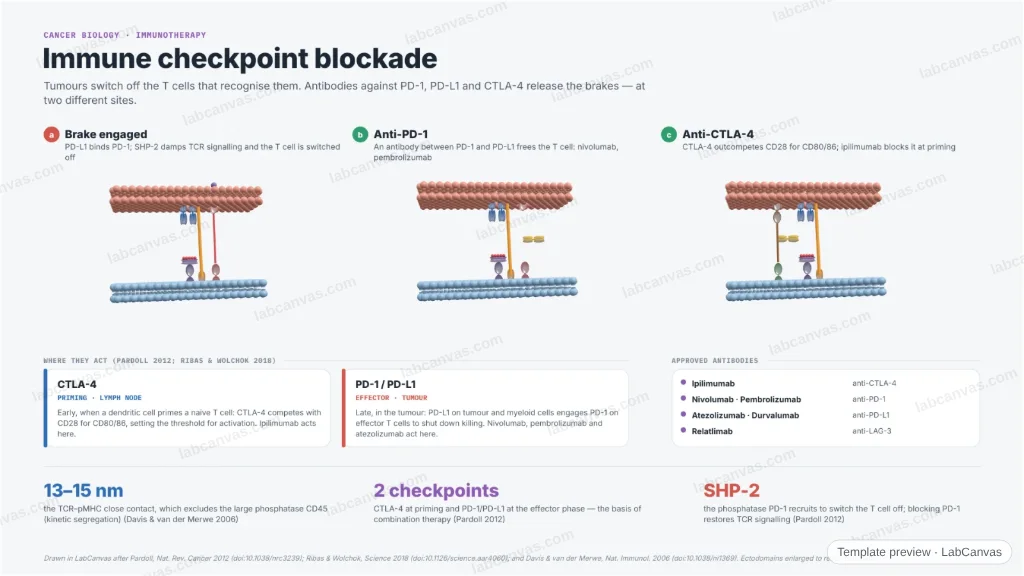
A figure of checkpoint blockade in cancer immunotherapy. The 3D immune-synapse model shows PD-1/PD-L1 engaged — recruiting SHP-2 to switch the T cell off — then an anti-PD-1 antibody and an anti-CTLA-4 antibody releasing the brake. It sets out where each checkpoint acts (CTLA-4 at priming in the lymph node, PD-1/PD-L1 at the effector phase in the tumour) and names the approved antibodies.
Updated 3 cited papers54 editable layers
At a glance
- Category
- Cancer biology
- Method
- Graphical abstract
- Product
- How checkpoint antibodies release the brakes on anti-tumour T cells
- Key result
- The TCR–pMHC close contact spans ~13–15 nm, excluding the large phosphatase CD45 (kinetic segregation) (Davis & van der Merwe 2006)
- Figure
- 1920 × 1080 px (16:9), 54 editable layers
Step by step
- Brake engagedPD-L1 on the tumour cell binds PD-1 on the T cell, which recruits the phosphatase SHP-2 to its ITIM/ITSM and damps TCR signalling (Pardoll 2012)
- Anti-PD-1 / anti-PD-L1An antibody wedged between PD-1 and PD-L1 frees the T cell; nivolumab and pembrolizumab block PD-1, atezolizumab blocks PD-L1 (Ribas & Wolchok 2018)
- Anti-CTLA-4CTLA-4 outcompetes CD28 for CD80/86 at priming; ipilimumab blocks CTLA-4 in the lymph node (Pardoll 2012)
- Two sites of actionCTLA-4 acts at T-cell priming in the lymph node; PD-1/PD-L1 acts at the effector phase in the tumour; LAG-3 is a third target (relatlimab) (Ribas & Wolchok 2018)
Uses
- Immuno-oncology review and graphical-abstract figures
- Talks on checkpoint inhibitors
- Teaching the two sites of checkpoint action
Papers this template follows
- Pardoll, “The blockade of immune checkpoints in cancer immunotherapy”, Nat. Rev. Cancer 2012. doi:10.1038/nrc3239review
- Ribas & Wolchok, “Cancer immunotherapy using checkpoint blockade”, Science 2018. doi:10.1126/science.aar4060review
- Davis & van der Merwe, “The kinetic-segregation model: TCR triggering and beyond”, Nat. Immunol. 2006. doi:10.1038/ni1369primary
Drawn in LabCanvas after Pardoll, Nat. Rev. Cancer 2012 (doi:10.1038/nrc3239); Ribas & Wolchok, Science 2018 (doi:10.1126/science.aar4060); and Davis & van der Merwe, Nat. Immunol. 2006 (doi:10.1038/ni1369). Ectodomains enlarged to read; schematic.
How templates are written and checked: editorial guidelines.
Questions
What does the “Immune checkpoint blockade” template show?
A figure of checkpoint blockade in cancer immunotherapy. The 3D immune-synapse model shows PD-1/PD-L1 engaged — recruiting SHP-2 to switch the T cell off — then an anti-PD-1 antibody and an anti-CTLA-4 antibody releasing the brake. It sets out where each checkpoint acts (CTLA-4 at priming in the lymph node, PD-1/PD-L1 at the effector phase in the tumour) and names the approved antibodies.
What are the steps shown in “Immune checkpoint blockade”?
1. Brake engaged: PD-L1 on the tumour cell binds PD-1 on the T cell, which recruits the phosphatase SHP-2 to its ITIM/ITSM and damps TCR signalling (Pardoll 2012). 2. Anti-PD-1 / anti-PD-L1: An antibody wedged between PD-1 and PD-L1 frees the T cell; nivolumab and pembrolizumab block PD-1, atezolizumab blocks PD-L1 (Ribas & Wolchok 2018). 3. Anti-CTLA-4: CTLA-4 outcompetes CD28 for CD80/86 at priming; ipilimumab blocks CTLA-4 in the lymph node (Pardoll 2012). 4. Two sites of action: CTLA-4 acts at T-cell priming in the lymph node; PD-1/PD-L1 acts at the effector phase in the tumour; LAG-3 is a third target (relatlimab) (Ribas & Wolchok 2018).
Which papers is “Immune checkpoint blockade” based on?
Pardoll, “The blockade of immune checkpoints in cancer immunotherapy”, Nat. Rev. Cancer 2012 (doi:10.1038/nrc3239); Ribas & Wolchok, “Cancer immunotherapy using checkpoint blockade”, Science 2018 (doi:10.1126/science.aar4060); Davis & van der Merwe, “The kinetic-segregation model: TCR triggering and beyond”, Nat. Immunol. 2006 (doi:10.1038/ni1369).
Can I edit the “Immune checkpoint blockade” template?
Yes. Open it in the LabCanvas editor: all 54 layers are editable. Change the text, colours and layout, then export PNG, TIFF or PDF at the width and DPI the journal asks for.
Do I need an account to use this template?
Not to view it here. To open and edit it, sign in to LabCanvas or create a free account (the free plan keeps up to 3 designs). Downloading the figure is part of LabCanvas Pro (US$19 a month or US$199 a year).