Graphical abstract template · Cancer biology · Graphical abstract
Nanomedicine and the EPR effect
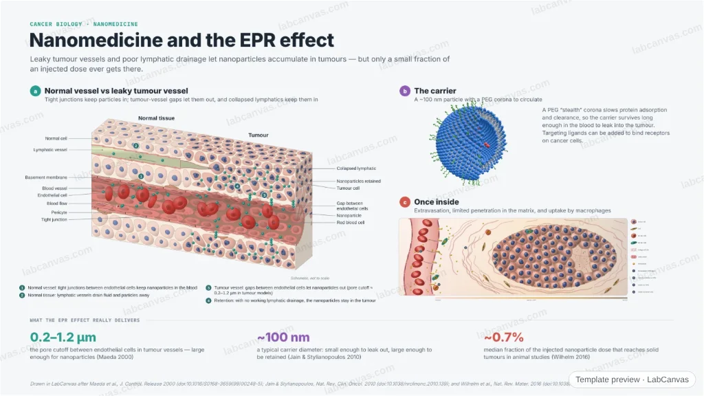
A figure of how nanomedicines reach a tumour. A 3D cutaway contrasts a normal vessel — tight junctions, draining lymphatics — with a leaky tumour vessel whose endothelial gaps let nanoparticles out, and whose collapsed lymphatics keep them in: the enhanced permeability and retention (EPR) effect. A PEGylated carrier and a 2D tumour scene show the reality — limited penetration and uptake by macrophages — and the delivery efficiency actually measured.
Updated 3 cited papers29 editable layers
At a glance
- Category
- Cancer biology
- Method
- Graphical abstract
- Product
- How nanoparticle carriers accumulate in a tumour, and why delivery is inefficient
- Key result
- Tumour-vessel pore cutoff ~0.2–1.2 µm; a median of ~0.7% of the injected nanoparticle dose reaches solid tumours in published animal studies (Wilhelm et al. 2016)
- Figure
- 1920 × 1080 px (16:9), 29 editable layers, 2 generated objects
Step by step
- Normal vesselTight junctions between endothelial cells keep nanoparticles in the blood; lymphatics drain fluid and particles away (Jain & Stylianopoulos 2010)
- The EPR effectLeaky tumour vessels have gaps between endothelial cells (pore cutoff ~0.2–1.2 µm) that let nanoparticles out; collapsed lymphatics keep them there (Maeda et al. 2000)
- The carrierA ~100 nm carrier with a PEG corona circulates long enough to reach the tumour; ligands can add active targeting
- The realityPenetration into dense matrix is limited and tumour-associated macrophages take up much of the dose; a median of only ~0.7% of the injected dose reaches the tumour (Wilhelm et al. 2016)
Uses
- Nanomedicine review and graphical-abstract figures
- Drug-delivery talks and posters
- Teaching passive targeting and its limits
Papers this template follows
- Maeda et al., “Tumor vascular permeability and the EPR effect in macromolecular therapeutics: a review”, J. Control. Release 2000. doi:10.1016/S0168-3659(99)00248-5method origin
- Jain & Stylianopoulos, “Delivering nanomedicine to solid tumors”, Nat. Rev. Clin. Oncol. 2010. doi:10.1038/nrclinonc.2010.139review
- Wilhelm et al., “Analysis of nanoparticle delivery to tumours”, Nat. Rev. Mater. 2016. doi:10.1038/natrevmats.2016.14primary
Drawn in LabCanvas after Maeda et al., J. Control. Release 2000 (doi:10.1016/S0168-3659(99)00248-5); Jain & Stylianopoulos, Nat. Rev. Clin. Oncol. 2010 (doi:10.1038/nrclinonc.2010.139); and Wilhelm et al., Nat. Rev. Mater. 2016 (doi:10.1038/natrevmats.2016.14). Schematic, not to scale.
How templates are written and checked: editorial guidelines.
Questions
What does the “Nanomedicine and the EPR effect” template show?
A figure of how nanomedicines reach a tumour. A 3D cutaway contrasts a normal vessel — tight junctions, draining lymphatics — with a leaky tumour vessel whose endothelial gaps let nanoparticles out, and whose collapsed lymphatics keep them in: the enhanced permeability and retention (EPR) effect. A PEGylated carrier and a 2D tumour scene show the reality — limited penetration and uptake by macrophages — and the delivery efficiency actually measured.
What are the steps shown in “Nanomedicine and the EPR effect”?
1. Normal vessel: Tight junctions between endothelial cells keep nanoparticles in the blood; lymphatics drain fluid and particles away (Jain & Stylianopoulos 2010). 2. The EPR effect: Leaky tumour vessels have gaps between endothelial cells (pore cutoff ~0.2–1.2 µm) that let nanoparticles out; collapsed lymphatics keep them there (Maeda et al. 2000). 3. The carrier: A ~100 nm carrier with a PEG corona circulates long enough to reach the tumour; ligands can add active targeting. 4. The reality: Penetration into dense matrix is limited and tumour-associated macrophages take up much of the dose; a median of only ~0.7% of the injected dose reaches the tumour (Wilhelm et al. 2016).
Which papers is “Nanomedicine and the EPR effect” based on?
Maeda et al., “Tumor vascular permeability and the EPR effect in macromolecular therapeutics: a review”, J. Control. Release 2000 (doi:10.1016/S0168-3659(99)00248-5); Jain & Stylianopoulos, “Delivering nanomedicine to solid tumors”, Nat. Rev. Clin. Oncol. 2010 (doi:10.1038/nrclinonc.2010.139); Wilhelm et al., “Analysis of nanoparticle delivery to tumours”, Nat. Rev. Mater. 2016 (doi:10.1038/natrevmats.2016.14).
Can I edit the “Nanomedicine and the EPR effect” template?
Yes. Open it in the LabCanvas editor: all 29 layers are editable, including 2 generated objects (structures, plots, equipment) with their own settings. Change the text, colours and layout, then export PNG, TIFF or PDF at the width and DPI the journal asks for.
Do I need an account to use this template?
Not to view it here. To open and edit it, sign in to LabCanvas or create a free account (the free plan keeps up to 3 designs). Downloading the figure is part of LabCanvas Pro (US$19 a month or US$199 a year).