Graphical abstract template · Cancer biology · Graphical abstract
The metastatic cascade
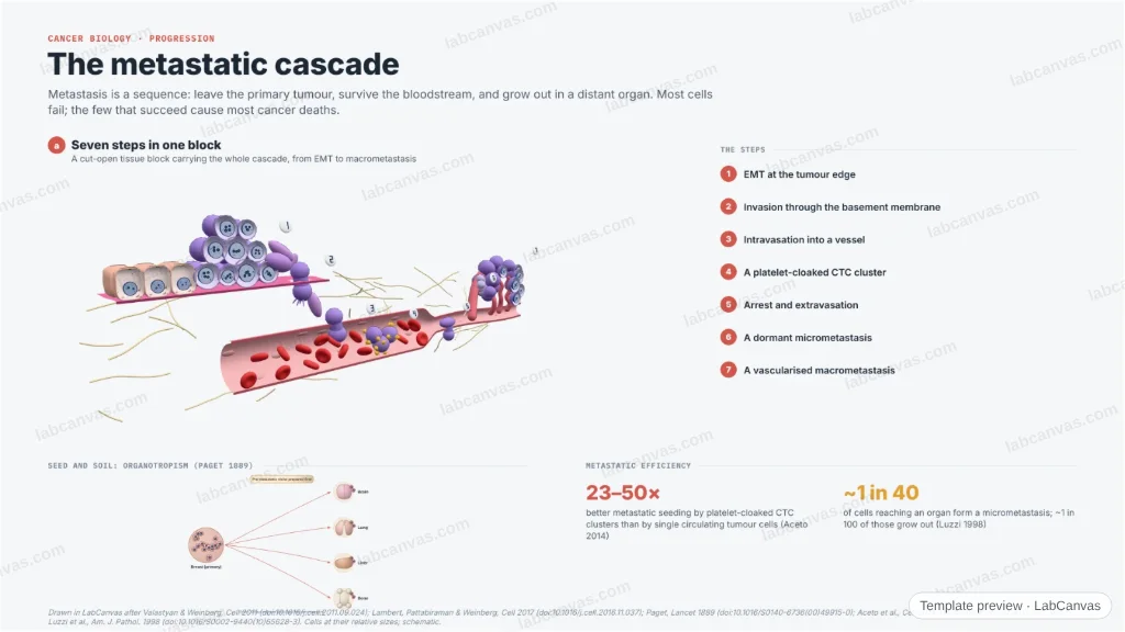
A figure of the invasion–metastasis cascade. A 3D cut-open block of tissue carries all seven steps — EMT, invasion through the basement membrane, intravasation, a platelet-cloaked CTC cluster, extravasation, a dormant micrometastasis and a vascularised macrometastasis — beside the seed-and-soil organotropism of breast cancer to bone, lung, liver and brain, with the pre-metastatic niche. Steps as in Valastyan & Weinberg 2011.
Updated 5 cited papers42 editable layers
At a glance
- Category
- Cancer biology
- Method
- Graphical abstract
- Product
- How a carcinoma cell leaves the primary tumour and colonises a distant organ
- Key result
- CTC clusters seed metastases 23–50× better than single CTCs (Aceto 2014); of cells reaching an organ, ~1 in 40 form micrometastases and ~1 in 100 of those grow out (Luzzi 1998)
- Figure
- 1920 × 1080 px (16:9), 42 editable layers, 1 generated objects
Step by step
- Local invasionEMT at the tumour edge; invadopodia breach the basement membrane into the stroma (Valastyan & Weinberg 2011)
- Intravasation & transitCells enter leaky vessels; CTC clusters cloaked in platelets survive the circulation and seed metastases far better than single cells (Aceto et al. 2014)
- Extravasation & dormancyCells arrest in a capillary, cross the wall and may lie dormant on the microvasculature for years (Valastyan & Weinberg 2011)
- ColonisationA few dormant cells outgrow into a vascularised macrometastasis; organotropism follows seed and soil (Paget 1889)
Uses
- Review and graphical-abstract figures on metastasis
- Talks on invasion and colonisation
- Teaching the invasion–metastasis cascade
Papers this template follows
- Valastyan & Weinberg, “Tumor metastasis: molecular insights and evolving paradigms”, Cell 2011. doi:10.1016/j.cell.2011.09.024review
- Lambert, Pattabiraman & Weinberg, “Emerging biological principles of metastasis”, Cell 2017. doi:10.1016/j.cell.2016.11.037review
- Paget, “The distribution of secondary growths in cancer of the breast”, Lancet 1889. doi:10.1016/S0140-6736(00)49915-0primary
- Aceto et al., “Circulating tumor cell clusters are oligoclonal precursors of breast cancer metastasis”, Cell 2014. doi:10.1016/j.cell.2014.07.013primary
- Luzzi et al., “Multistep nature of metastatic inefficiency”, Am. J. Pathol. 1998. doi:10.1016/S0002-9440(10)65628-3primary
Drawn in LabCanvas after Valastyan & Weinberg, Cell 2011 (doi:10.1016/j.cell.2011.09.024); Lambert, Pattabiraman & Weinberg, Cell 2017 (doi:10.1016/j.cell.2016.11.037); Paget, Lancet 1889 (doi:10.1016/S0140-6736(00)49915-0); Aceto et al., Cell 2014 (doi:10.1016/j.cell.2014.07.013); and Luzzi et al., Am. J. Pathol. 1998 (doi:10.1016/S0002-9440(10)65628-3). Cells at their relative sizes; schematic.
How templates are written and checked: editorial guidelines.
Questions
What does the “The metastatic cascade” template show?
A figure of the invasion–metastasis cascade. A 3D cut-open block of tissue carries all seven steps — EMT, invasion through the basement membrane, intravasation, a platelet-cloaked CTC cluster, extravasation, a dormant micrometastasis and a vascularised macrometastasis — beside the seed-and-soil organotropism of breast cancer to bone, lung, liver and brain, with the pre-metastatic niche. Steps as in Valastyan & Weinberg 2011.
What are the steps shown in “The metastatic cascade”?
1. Local invasion: EMT at the tumour edge; invadopodia breach the basement membrane into the stroma (Valastyan & Weinberg 2011). 2. Intravasation & transit: Cells enter leaky vessels; CTC clusters cloaked in platelets survive the circulation and seed metastases far better than single cells (Aceto et al. 2014). 3. Extravasation & dormancy: Cells arrest in a capillary, cross the wall and may lie dormant on the microvasculature for years (Valastyan & Weinberg 2011). 4. Colonisation: A few dormant cells outgrow into a vascularised macrometastasis; organotropism follows seed and soil (Paget 1889).
Which papers is “The metastatic cascade” based on?
Valastyan & Weinberg, “Tumor metastasis: molecular insights and evolving paradigms”, Cell 2011 (doi:10.1016/j.cell.2011.09.024); Lambert, Pattabiraman & Weinberg, “Emerging biological principles of metastasis”, Cell 2017 (doi:10.1016/j.cell.2016.11.037); Paget, “The distribution of secondary growths in cancer of the breast”, Lancet 1889 (doi:10.1016/S0140-6736(00)49915-0); Aceto et al., “Circulating tumor cell clusters are oligoclonal precursors of breast cancer metastasis”, Cell 2014 (doi:10.1016/j.cell.2014.07.013); Luzzi et al., “Multistep nature of metastatic inefficiency”, Am. J. Pathol. 1998 (doi:10.1016/S0002-9440(10)65628-3).
Can I edit the “The metastatic cascade” template?
Yes. Open it in the LabCanvas editor: all 42 layers are editable, including 1 generated objects (structures, plots, equipment) with their own settings. Change the text, colours and layout, then export PNG, TIFF or PDF at the width and DPI the journal asks for.
Do I need an account to use this template?
Not to view it here. To open and edit it, sign in to LabCanvas or create a free account (the free plan keeps up to 3 designs). Downloading the figure is part of LabCanvas Pro (US$19 a month or US$199 a year).