Graphical abstract template · Cancer biology · Graphical abstract
Liquid biopsy
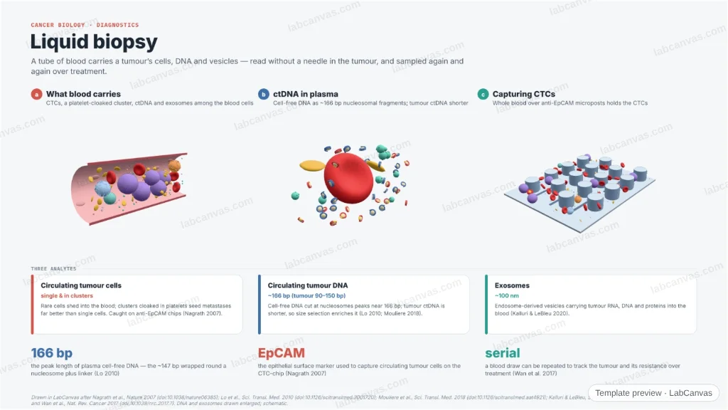
A figure of what a blood sample reveals about a tumour. Three 3D scenes show the analytes of a liquid biopsy — circulating tumour cells (single and in platelet-cloaked clusters), circulating tumour DNA as nucleosome-sized fragments, and exosomes — and how CTCs are captured on an anti-EpCAM microchip. The three analytes and their real sizes are set out alongside.
Updated 5 cited papers43 editable layers
At a glance
- Category
- Cancer biology
- Method
- Graphical abstract
- Product
- Tumour analytes read from a blood draw: CTCs, ctDNA and exosomes
- Key result
- cfDNA peaks at ~166 bp (Lo 2010); tumour ctDNA enriched at ~90–150 bp (Mouliere 2018); exosomes ~100 nm (Kalluri & LeBleu 2020); the CTC-chip runs whole blood over ~78,000 anti-EpCAM microposts (Nagrath 2007)
- Figure
- 1920 × 1080 px (16:9), 43 editable layers
Step by step
- What blood carriesCirculating tumour cells (single and in platelet-cloaked clusters), cell-free and tumour DNA, and exosomes travel in the plasma among the blood cells (Wan et al. 2017)
- ctDNACell-free DNA is cut like nucleosomes and peaks at ~166 bp; tumour-derived ctDNA is shorter, enriched at ~90–150 bp (Lo et al. 2010; Mouliere et al. 2018)
- ExosomesEndosome-derived vesicles about 100 nm across carry tumour nucleic acids and proteins (Kalluri & LeBleu 2020)
- Capturing CTCsCTCs are caught from whole blood on anti-EpCAM-coated microposts while blood cells flow past (Nagrath et al. 2007)
Uses
- Diagnostics review and graphical-abstract figures
- Talks on circulating biomarkers
- Teaching the analytes of a liquid biopsy
Papers this template follows
- Nagrath et al., “Isolation of rare circulating tumour cells in cancer patients by microchip technology”, Nature 2007. doi:10.1038/nature06385primary
- Lo et al., “Maternal plasma DNA sequencing reveals the genome-wide genetic and mutational profile of the fetus”, Sci. Transl. Med. 2010. doi:10.1126/scitranslmed.3001720primary
- Mouliere et al., “Enhanced detection of circulating tumor DNA by fragment size analysis”, Sci. Transl. Med. 2018. doi:10.1126/scitranslmed.aat4921primary
- Kalluri & LeBleu, “The biology, function, and biomedical applications of exosomes”, Science 2020. doi:10.1126/science.aau6977review
- Wan et al., “Liquid biopsies come of age: towards implementation of circulating tumour DNA”, Nat. Rev. Cancer 2017. doi:10.1038/nrc.2017.7review
Drawn in LabCanvas after Nagrath et al., Nature 2007 (doi:10.1038/nature06385); Lo et al., Sci. Transl. Med. 2010 (doi:10.1126/scitranslmed.3001720); Mouliere et al., Sci. Transl. Med. 2018 (doi:10.1126/scitranslmed.aat4921); Kalluri & LeBleu, Science 2020 (doi:10.1126/science.aau6977); and Wan et al., Nat. Rev. Cancer 2017 (doi:10.1038/nrc.2017.7). DNA and exosomes drawn enlarged; schematic.
How templates are written and checked: editorial guidelines.
Questions
What does the “Liquid biopsy” template show?
A figure of what a blood sample reveals about a tumour. Three 3D scenes show the analytes of a liquid biopsy — circulating tumour cells (single and in platelet-cloaked clusters), circulating tumour DNA as nucleosome-sized fragments, and exosomes — and how CTCs are captured on an anti-EpCAM microchip. The three analytes and their real sizes are set out alongside.
What are the steps shown in “Liquid biopsy”?
1. What blood carries: Circulating tumour cells (single and in platelet-cloaked clusters), cell-free and tumour DNA, and exosomes travel in the plasma among the blood cells (Wan et al. 2017). 2. ctDNA: Cell-free DNA is cut like nucleosomes and peaks at ~166 bp; tumour-derived ctDNA is shorter, enriched at ~90–150 bp (Lo et al. 2010; Mouliere et al. 2018). 3. Exosomes: Endosome-derived vesicles about 100 nm across carry tumour nucleic acids and proteins (Kalluri & LeBleu 2020). 4. Capturing CTCs: CTCs are caught from whole blood on anti-EpCAM-coated microposts while blood cells flow past (Nagrath et al. 2007).
Which papers is “Liquid biopsy” based on?
Nagrath et al., “Isolation of rare circulating tumour cells in cancer patients by microchip technology”, Nature 2007 (doi:10.1038/nature06385); Lo et al., “Maternal plasma DNA sequencing reveals the genome-wide genetic and mutational profile of the fetus”, Sci. Transl. Med. 2010 (doi:10.1126/scitranslmed.3001720); Mouliere et al., “Enhanced detection of circulating tumor DNA by fragment size analysis”, Sci. Transl. Med. 2018 (doi:10.1126/scitranslmed.aat4921); Kalluri & LeBleu, “The biology, function, and biomedical applications of exosomes”, Science 2020 (doi:10.1126/science.aau6977); Wan et al., “Liquid biopsies come of age: towards implementation of circulating tumour DNA”, Nat. Rev. Cancer 2017 (doi:10.1038/nrc.2017.7).
Can I edit the “Liquid biopsy” template?
Yes. Open it in the LabCanvas editor: all 43 layers are editable. Change the text, colours and layout, then export PNG, TIFF or PDF at the width and DPI the journal asks for.
Do I need an account to use this template?
Not to view it here. To open and edit it, sign in to LabCanvas or create a free account (the free plan keeps up to 3 designs). Downloading the figure is part of LabCanvas Pro (US$19 a month or US$199 a year).